
FIGURE
Fig. 7
- ID
- ZDB-FIG-071112-8
- Publication
- Kosaka et al., 2007 - Spatiotemporal localization of germ plasm RNAs during zebrafish oogenesis
- Other Figures
- All Figure Page
- Back to All Figure Page
Fig. 7
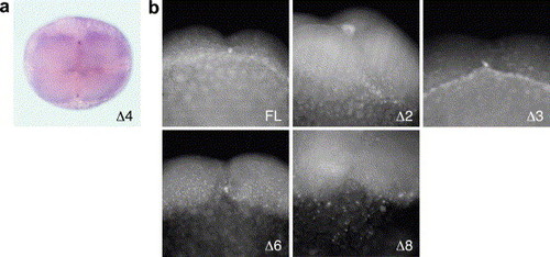
Distribution of a reporter mRNA fused with mutant dazl 3′ UTRs in 4-cell embryos. (a) In situ hybridization of Δ4 RNA. Animal view of the 4-cell embryo shown in Fig. 6b. (b) Fluorescein in situ hybridization in transgenic 4-cell embryos expressing GFP mRNA fused with full-length (FL), Δ2, Δ3, Δ6, or Δ8 dazl 3′ UTRs. The furrow region is enlarged. |
Expression Data
Expression Detail
Antibody Labeling
Phenotype Data
Phenotype Detail
Acknowledgments
This image is the copyrighted work of the attributed author or publisher, and
ZFIN has permission only to display this image to its users.
Additional permissions should be obtained from the applicable author or publisher of the image.
Reprinted from Mechanisms of Development, 124(4), Kosaka, K., Kawakami, K., Sakamoto, H., and Inoue, K., Spatiotemporal localization of germ plasm RNAs during zebrafish oogenesis, 279-289, Copyright (2007) with permission from Elsevier. Full text @ Mech. Dev.