
Fig. 2
- ID
- ZDB-FIG-070822-51
- Publication
- Hinits et al., 2007 - Mrf4 (myf6) is dynamically expressed in differentiated zebrafish skeletal muscle
- Other Figures
- All Figure Page
- Back to All Figure Page
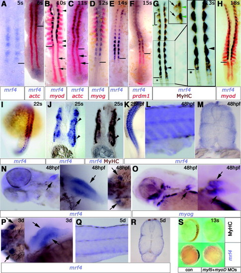
Mrf4 mRNA is expressed in terminally differentiated skeletal muscle. In situ mRNA hybridisation for mrf4 (purple, A–N, P, Q , R, S), myoD (red, B,H), myogenin (myog red, D; purple, O) actin (actc red, A,C) prdm1 (red, F) and wholemount immunohistochemistry for MyHC (brown, G, J, S) in zebrafish embryos, viewed in dorsal flatmount (A–H), wholemount (K, L, N centre; O, Q, S lateral view; I, N right; P left dorsal view; N left ventral view) or transverse cryosection (J, M, R). Anterior is to the top (A–I, K, S), or left (L, N–Q) and dorsal to top (M,R). (A) Mrf4 is first detected adaxially in each somite at 5 and 6s, after actin (right panel). The horizontal line indicates the newest somite border (A–H). (B–D) At 10–12s, mrf4 mRNA is restricted to somitic adaxial cells, whereas myoD (B) and actin (C) expression are strong in adaxial cells of presomitic mesoderm (arrows) and myod (B) and myog (D) in the lateral fast muscle precursors (arrowheads). Myogenin mRNA (D) shows that mrf4 is not expressed in fast precursors at this stage. Dual immunohistochemical detection of two mRNAs in a single embryo reveals expression of mrf4 exclusively in the adaxial location of slow muscle obscuring the red myod or actin label. Note that myogenin expression is not detected in presomitic adaxial cells (Weinberg et al., 1996) due to short incubation with substrate. (E–H) Mrf4 is down-regulated in rostral somites before 14s (E). Mrf4 and prdm1 mRNAs are co-expressed in differentiated adaxial cells whereas prdm1 alone is expressed in presomitic adaxial cells (F). Mrf4 transcripts co-localise with MyHC protein in adaxial cells of the newly forming somite (G, asterisks). Mrf4 expression is strongest in the six most recently formed somites, obscuring the MyHC signal (G, arrowheads; box shown magnified at right). In more mature somites, declining mrf4 is detected in a similar location (G, bracketed region shown in inset, green arrowheads), whereas MyHC-expressing slow fibres are increasing (G inset, arrows). By 18s, mrf4 is expressed most strongly in six nascent somites (H). (I) At 22s, mrf4 is up-regulated once more in rostral somites. (J) By 25s, mrf4 mRNA is present throughout the somite and at high level in slow fibres after their migration (arrowheads). Re-staining of the same section shows that slow MyHC (F59) overlaps the high mrf4 expressing cells (arrowheads, right panel). (K) At 26hpf, mrf4 is broadly expressed in muscle. (L and M) Mrf4 expression at 48hpf appears stronger in the superficial slow muscle cells. (N and O) Mrf4 and myogenin are expressed in dorsal and ventral muscle masses of pectoral fin at 48hpf (arrows). High magnifications of the fin show mrf4 and myogenin expression in dorsal and ventral muscle masses. Myogenin is detected in numerous ventral head muscle anlage (O left), but mrf4 mRNA shows no such pattern (N left). (P) Mrf4 mRNA persists in pectoral fin at 72hpf. (Q and R) Mrf4 is weakly expressed at 5d and is more readily detected superficial slow cells. (S) Embryos injected with myf5 and myoD antisense morpholinos have no MyHC and lack mrf4 expression at 12s. |
| Genes: | |
|---|---|
| Fish: | |
| Knockdown Reagents: | |
| Anatomical Terms: | |
| Stage Range: | 5-9 somites to Day 5 |
Reprinted from Gene expression patterns : GEP, 7(7), Hinits, Y., Osborn, D.P., Carvajal, J.J., Rigby, P.W., and Hughes, S.M., Mrf4 (myf6) is dynamically expressed in differentiated zebrafish skeletal muscle, 738-745, Copyright (2007) with permission from Elsevier. Full text @ Gene Expr. Patterns