
Fig. 4
- ID
- ZDB-FIG-070821-7
- Publication
- Wallace et al., 2005 - Intestinal growth and differentiation in zebrafish
- Other Figures
- All Figure Page
- Back to All Figure Page
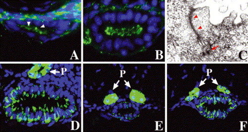
Step-wise polarization of developing intestinal epithelia: (A) cross-section through the anterior intestine of a 50 hpf embryo processed for ZO-1 immunohistochemistry. Apical ZO-1 (arrowheads) is first evident at this stage. (B) Apical ZO-1 staining in the anterior intestine is pronounced at 74 hpf. (C) Transmission electron micrograph (TEM) shows well-defined tight junction (arrowheads) and desmosomes (arrow) in an intestial epithelial cell of a 74 hpf larva. (D–F) Basolateral Na/K–ATPase is present in the 74 hpf anterior intestinal epithelium, but there is minimal staining in the posterior intestine at this stage (E). Pronounced Na/K–ATPase is present in the posterior intestine at 5 dpf (F). (P) Pronephric ducts. |
Reprinted from Mechanisms of Development, 122(2), Wallace, K.N., Akhter, S., Smith, E.M., Lorent, K., and Pack, M., Intestinal growth and differentiation in zebrafish, 157-73, Copyright (2005) with permission from Elsevier. Full text @ Mech. Dev.