
Fig. 1
- ID
- ZDB-FIG-070821-5
- Publication
- Wallace et al., 2005 - Intestinal growth and differentiation in zebrafish
- Other Figures
- All Figure Page
- Back to All Figure Page
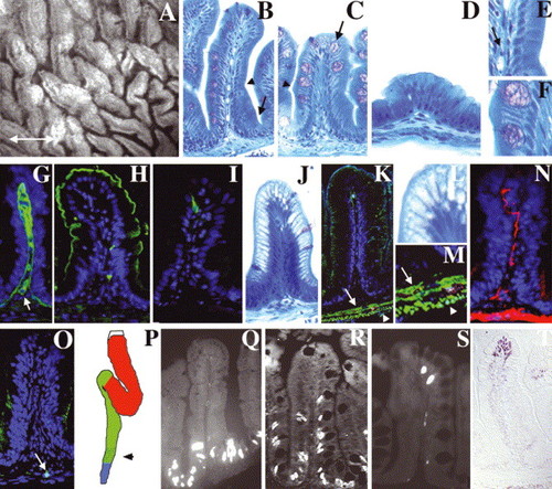
Adult zebrafish intestine: (A) Whole-mount, interior view of the adult zebrafish anterior intestine reveals irregularly shaped epithelial folds. The long axis of most folds is oriented circumferentially; others are oriented randomly (double arrow defines anterior–posterior axis). (B–D) Histological cross-section through the folds of the anterior (B), mid (C) and posterior (D) intestine. Note progressive diminution of the height of mid and posterior folds. A connective tissue core containing blood vessels and nerves underlies the epithelium of each fold. Note absence of crypts at base of folds (arrow in B). (E,F) Enlargement of region adjacent to arrowheads in (B,C) showing columnar-shaped enterocytes and goblet cells. Arrow in (E) points to an erythrocyte in a blood vessel in the connective tissue of the intestinal fold (G) Laminin immunohistochemistry identifies the basement membrane separating the epithelium from the underlying connective tissue (lamina propria). Laminin also encircles blood vessels within the lamina propria (arrow). Four principal cell types are identified within the epithelium. (H) Enterocytes are identified by apical sodium phosphate transporter (NaPi). (I) Enteroendocrine cells are identified by pancreatic polypeptide. (C,F) Goblet cells are identified histologically by their mucinous theca (arrow in C). (J,L) Antigen presenting cells within segment II of the mid-intestine are identified by their distinctive supra-nuclear vacuole. (K,M) Desmin (green) identifies inner circular (arrow) and outer longitudinal (arrowhead) smooth muscle layers beneath the lamina propria. (N) Acetylated tubulin identifies axonal fibers within the muscularis and the lamina propria. (O) Hu immunohistochemistry identifies a solitary enteric neuron cell body within the muscularis (arrow). (P) Schematic representing the anterior (red), mid (green) and posterior (blue) intestinal segments. Stippled region of the mid segment represents location of specialized enterocytes. (Q–S) Intestinal epithelial cell renewal revealed by BrdU immunohistochemistry. S-phase cells are present at the base of the intestinal folds 1 h after BrdU injection (Q). 4-day post-injection, S-phase cells are identified at the base and in the mid fold region (R). 7-day post-injection, S-phase cells are only identified at the tip of the folds (S). (T) TUNEL assay identifies apoptotic cells at the tips of intestinal folds. |
Reprinted from Mechanisms of Development, 122(2), Wallace, K.N., Akhter, S., Smith, E.M., Lorent, K., and Pack, M., Intestinal growth and differentiation in zebrafish, 157-73, Copyright (2005) with permission from Elsevier. Full text @ Mech. Dev.