
Fig. 3
- ID
- ZDB-FIG-070821-6
- Publication
- Wallace et al., 2005 - Intestinal growth and differentiation in zebrafish
- Other Figures
- All Figure Page
- Back to All Figure Page
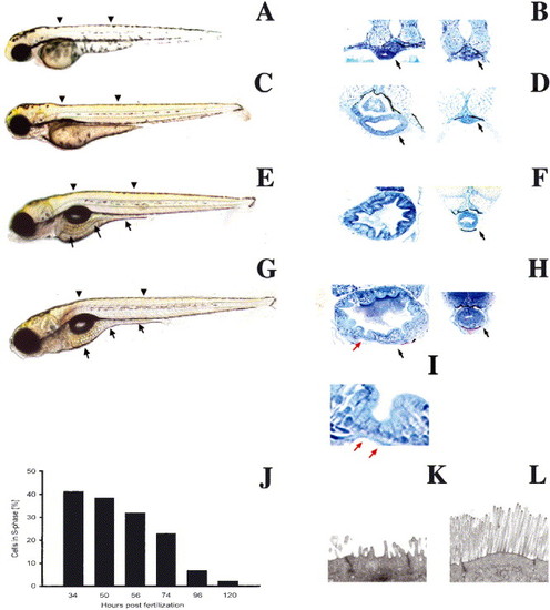
Biphasic intestinal growth in zebrafish embryos and larvae: (A–D) Lateral views of 50, 74, 96 and 120 hpf zebrafish. The intestine is prominent at 96 and 120 hpf (arrows C, D) but difficult to identify at 50 (A) and 72 hpf (B). (E–H) Corresponding histological cross-sections through the anterior (left) and posterior (right) intestine of zebrafish depicted in (A–D). Note appearance of folds and columnar-shaped epithelial cells between 72 and 96 hpf. Folds are cut in cross-section (red arrow) and tangential to the long fold axis (black arrow). (I) High power view of intestinal folds in H shows cuboidal cells at base of fold (red arrow) and adjacent columnar cells. (J) Epithelial cell proliferation is biphasic; nadir is reached at 120 hpf and cell proliferation does not increase within the first 24 hpf of feeding (not shown). Arrowheads identify levels of histological sections. Arrows in histological sections point to the intestine unless otherwise noted. (K,L) Transmission electron micrographs showing the apical region of a 74 (K) and 120 hpf (L) intestinal epithelial cell. |
Reprinted from Mechanisms of Development, 122(2), Wallace, K.N., Akhter, S., Smith, E.M., Lorent, K., and Pack, M., Intestinal growth and differentiation in zebrafish, 157-73, Copyright (2005) with permission from Elsevier. Full text @ Mech. Dev.