|
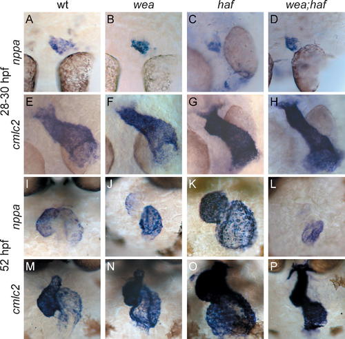
Regionalization of nppa Expression in wea, haf, and wea;haf Mutants Is Normal at LHT Stages but Abnormal at Expanded Chamber Stages. Whole-mount in situ hybridization of nppa (A–D and I–L) compared to cmlc2 (E–H and M–P). (A–H) Dorsal views, ventricle to the top, at LHT stages. (A, B, E, and F) are 28 hpf; (C, D, G, and H) are 30 hpf. Although initiation of nppa expression is delayed in haf (C) and wea;haf (D) mutants, the nppa expression domain is normal in the wea (B), haf (C), and wea;haf (D) LHT. (I–P) Frontal views, ventricle to the left, at expanded chamber stages (52 hpf). (J) Regionalized nppa expression is not maintained in the wea mutant ventricle; instead, nppa expression in the wea ventricle is fainter and in a smaller domain than in wild type (wt) (I). (K) In the haf mutant ventricle, intense nppa expression is found throughout the chamber, rather than being restricted to the OC. (L) The wea;haf double mutant ventricle exhibits weak and diffuse nppa expression.
|