|
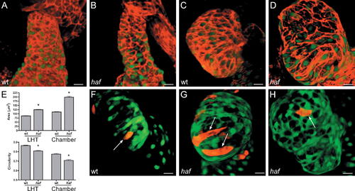
Cardiomyocyte Surfaces throughout the haf Ventricle Become Excessively Enlarged and Elongated. (A–D) Phalloidin staining (red) of wt and haf mutant hearts expressing Tg(cmlc2:egfp) at LHT (A and B) and expanded chamber (C and D) stages. (E) Bar graphs depict surface area and circularity measurements, as in Figure 2. An asterisk indicates statistically significant differences compared to wild-type (wt) data (p < 0.0001). The shape and size of haf cells are significantly different from those of wt cells; at both LHT and expanded chamber stages, haf cells are larger and more elongated. (F–H) Confocal projections of live hearts, as in Figure 2, confirm the abnormal size and shape of haf mutant cardiomyocytes in the expanded ventricle. haf cells ([G and H] arrows) are larger than their wt counterparts ([F] arrow) and can be greatly elongated (G). Size bar represents 20 μm.
|