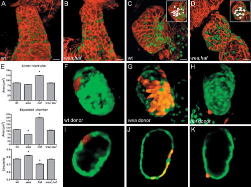
Cells Lacking Vmhc Assume Different Morphologies under Different Hemodynamic Conditions. (A–D) Phalloidin staining (red) of wild-type (wt) and wea;haf double mutant hearts expressing Tg(cmlc2:egfp) at LHT (A and B) and expanded chamber (C and D) stages. Insets highlight representative cell shapes in white. wea;haf cells are irregularly shaped and lack apparent organization or alignment. (E) Bar graphs depict surface area and circularity measurements, as in Figure 2. An asterisk indicates statistically significant differences compared to wt data (p < 0.0001). Despite their irregular organization and morphology, wea;haf ventricular cells exhibit a size and circularity range similar to that of wt cells and distinct from that of wea or haf. Notably, wea;haf ventricular cells enlarge more than wea cells do (p < 0.0001), even though wea;haf mutants lack blood flow. Also, wea;haf cell morphologies are not as extreme in size (p < 0.0001) or elongation (p < 0.001) as haf cell morphologies, even though wea;haf mutant cells lack Vmhc. (F–K) Chimeric ventricles resulting from transplantation of rhodamine dextran-labeled blastomeres into wt hosts expressing Tg(cmlc2:egfp). Optical sections of (F, G, and H) are shown in (I, J, and K), respectively. (F, G, I, and J) Cells from wt or wea donors integrate normally within the wall of the wt host ventricle. (H and K) In contrast, cells from haf donors project abnormally from the ventricular wall and fail to maintain normal shape, indicating a cell-autonomous requirement for Vmhc in the maintenance of cardiomyocyte morphology. Size bar represents 20 μm.
|
