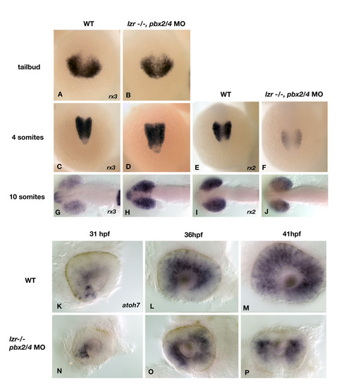
The expression of rx2, rx3, and atoh7 is aberrant in pbx2/4 null embryos. The expression of rx3, the earliest known eye field marker is unaffected at the tailbud and 4 somite stage (A-D) but is increased in expression at the 10 somite stage (H) when compared to wildtype (G). The expression of rx2 is reduced at both the 4 and 10 somite stage in pbx2/4 null embryos (F and J), when compared to wildtype (E and I). No expression of rx2 is observed at the tailbud stage. The expression of atoh7 was analyzed as a marker of differentiating retinal ganglion cells. In wild type embryos, expression begins in the ventral nasal domain of the retina at about 31 hpf (K). Expression proceeds in a wave-like fashion to include the dorsal retina by 36 hpf (L), and has filled the entire retina by 41 hpf (M). In pbx2/4 null embryos, expression is initiated correctly at 31 hpf (N). Expression proceeds in a wave-like fashion but is excluded from the dorsal domain of the retina at both 36 hpf (O) and 41 hpf (P).
|
