
Fig. 3
- ID
- ZDB-FIG-070815-21
- Publication
- Zhao et al., 2007 - Genetic defects of pronephric cilia in zebrafish
- Other Figures
- All Figure Page
- Back to All Figure Page
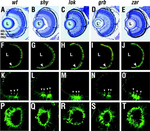
Phenotype of sensory neurons. (A–E) Transverse plastic sections through the central retina near the optic nerve at 7 dpf. (F–J) Confocal images of transverse frozen sections through the retina near the optic nerve stained with Zpr1 (green) and anti-blue opsin (red) antibodies at 7 dpf. (K–O) Anterior tether cells (arrowheads) in otic vesicles immunostained for acetylated-α-tubulin at 30 hpf. (P–T) Nose cilia stained for acetylated-α-tubulin at 3 dpf. In A–J, “L” indicates the lens, midline is right, dorsal is up. Arrowheads in (F–J) indicate the photoreceptor cell layer. GCL, ganglion cell layer; INL, inner nuclear layer; PRCL, photoreceptor cell layer. |
| Fish: | |
|---|---|
| Observed In: | |
| Stage: | Days 7-13 |
Reprinted from Mechanisms of Development, 124(7-8), Zhao, C., and Malicki, J., Genetic defects of pronephric cilia in zebrafish, 605-616, Copyright (2007) with permission from Elsevier. Full text @ Mech. Dev.