
FIGURE
Fig. 1
- ID
- ZDB-FIG-070815-19
- Publication
- Zhao et al., 2007 - Genetic defects of pronephric cilia in zebrafish
- Other Figures
- All Figure Page
- Back to All Figure Page
Fig. 1
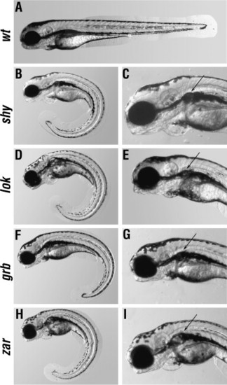
External phenotypes of mutant animals. Lateral views of wild-type (A) and mutant (B–I) larvae at 3 dpf. (C, E, G, and I), show high magnifications of the anterior region of mutant larvae. All mutants shown develop kidney cysts (arrows in C, E, G, and I) and ventrally curled tails (B, D, F, and H). There are no obvious differences in eye and ear phenotypes between mutant and wild type larvae. In all panels, anterior is left, dorsal is up. |
Expression Data
Expression Detail
Antibody Labeling
Phenotype Data
| Fish: | |
|---|---|
| Observed In: | |
| Stage: | Protruding-mouth |
Phenotype Detail
Acknowledgments
This image is the copyrighted work of the attributed author or publisher, and
ZFIN has permission only to display this image to its users.
Additional permissions should be obtained from the applicable author or publisher of the image.
Reprinted from Mechanisms of Development, 124(7-8), Zhao, C., and Malicki, J., Genetic defects of pronephric cilia in zebrafish, 605-616, Copyright (2007) with permission from Elsevier. Full text @ Mech. Dev.