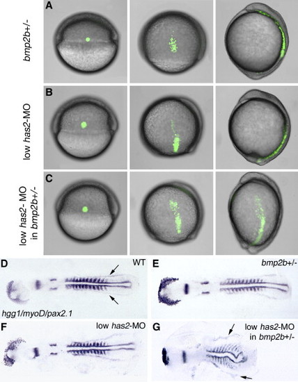
bmp2b and has2 Display Genetic Interaction during Dorsal Convergence, but Not Dorsoventral Patterning. (A–C) Live embryos with fluorescently labeled group of lateral mesodermal cells after treatment indicated on the left side of each row, at the onset, the middle, and the end of gastrulation; lateral views, dorsal to the right, anterior at the top. For wild-type controls, see Figure 1B. (D–G) Same embryos after whole-mount in situ hybridization at the 12-somite stage with hgg1, marking hatching gland precursor cells, myoD, marking muscle precursors, and pax2.1, marking midbrain-hindbrain boundary, otic placodes, and pronephric ducts. Note that in (G), the bmp2b heterozygote injected with low amounts of has2 MO displays an undulated notochord and laterally expanded somites, resulting from reduced convergence movements, while pax2.1-positive pronephric cells (indicated by arrows in [D] and [G]) are present in normal numbers, indicating that the embryo is not dorsalized (compare with [3]).
|
