|
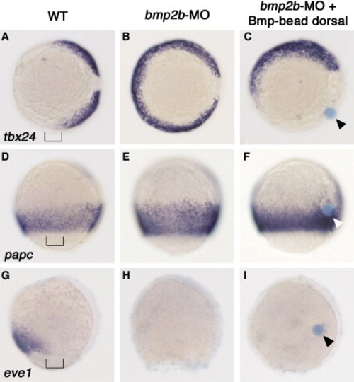
The Bmp Beads Generate a Broad Field of Laterally Specified Mesoderm. Embryos at the 80% epiboly stage, dorsal to the right, after whole-mount in situ hybridizations with probes indicated on the left side of each row, and after treatment indicated on the top of each column. Vegetal view shown in (A)–(C); lateral view shown in (D)–(I), anterior to the top. Bmp beads are indicated with arrowheads. The mesoderm throughout the entire bead-bearing side of the embryos displays absence of tbx24 and eve1, but presence of papc [46] transcripts, as is normally characteristic for a narrow lateral domain of wild-type (WT) embryos at 90° from the shield (indicated by brackets in [A], [D], and [G]). Note the presence of papc-positive cells in close proximity to the bead in (F), in line with the observation that cell movements become prominent only after mid-gastrula stages.
|