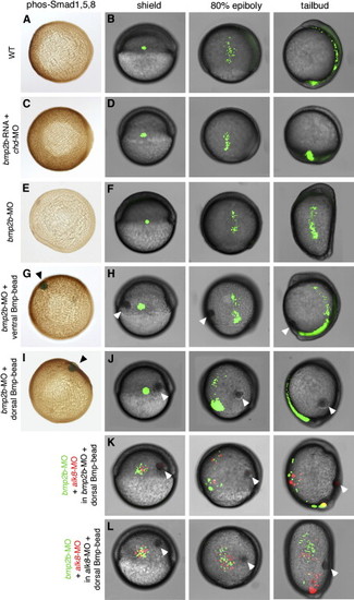
The Bmp Gradient Determines the Direction of Cell Movements during Dorsal Convergence of the Lateral Mesoderm in a Non-Cell-Autonomous Fashion (A, C, E, G, and I) Vegetal views of embryos at 85% epiboly after anti-phospho Smad1/5 immunostainings, dorsal to the right. (B, D, F, H, J–L) All other panels show live embryos after photoactivation of caged fluorescent dextran in a group of cells in the lateral mesoderm (B, D, F, H, and J) or after transplantation of a mixture of lateral mesodermal cells containing fluorescein or rhodamin dextran (K and L). Each embryo is shown at shield stage (early gastrula, 6 hpf), at 80% epiboly stage (mid-gastrula, 8 hpf), and at tailbud stage (end of gastrulation, 10 hpf). Dorsal is to the right, anterior/animal pole at the top. Embryos were injected either with bmp2b mRNA and chordin MO (C and D), with bmp2b MO (E–K), or with alk8 MO (L). To restore or generate a reverse Bmp gradient, Bmp beads (marked with arrowheads) were implanted into ventrolateral (G and H) or dorsolateral (I–L) positions of bmp2b or alk8 morphants at shield stage.
|
