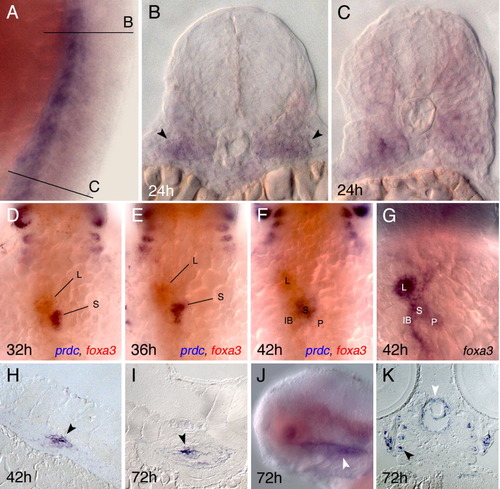
Prdc expression in somites, swim bladder, and oral epithelium. A-C: prdc is expressed primarily in the ventral part of the developing somites at 24 hours postfertilization (hpf; arrowheads). B and C are plastic sections of the whole-mount prdc in situ hybridization shown in A. The section planes are marked with the lines B and C. D-F: prdc (in blue) and foxa3 (in red) expression at 32, 36, and 42 hpf (D-F). The two probes overlap at the site of the swim bladder bud. The prdc expression in pharyngeal arches is also visible at the top. G: Single foxa3 staining marks the liver (L), swim bladder (S), pancreas (P), and buds and the intestinal bulb (IB). H,I: sections of prdc probe-stained embryos depict expression in the swim bladder mesenchyme (arrowheads) at 42 and 72 hpf. J,K: prdc expression at 72 hpf marks the oral epithelium in whole-mount staining and sections (white arrowheads). Staining in the arches is also seen (black arrowhead in K). Anterior is to the top in panels A, D-G and to the left in J. h, hours postfertilization.
|
