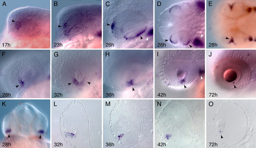
Expression of prdc in the developing eye. A,B: Prdc transcripts appear at 17 hours postfertilization (hpf) in the anterodorsal region of the developing optic lobe and remain in this site till around 23 hpf (black arrowheads). C,D: prdc expression marks the dorsal and ventral outer edges of the optic cup at 26 hpf (black arrowheads). D is dorsal view of C. White arrowheads point to prdc expression in the first pharyngeal arch. E-J: prdc expression in the retinal epithelium (black arrowheads) is localized ventrally at the edges surrounding the choroid fissure (28-42 hpf). E is dorsal view of F. J: At 72 hpf, prdc expression can be seen in a very small ventral region in the marginal zone. K-O: Frontal view (K) of prdc staining in the retina at 28 hpf and sections (L-O) of the corresponding whole-mounts in the adjacent top panels at 32, 36, 42, and 72 hpf illustrate that, after the eye rotates, prdc expression is confined to ventral areas of the developing retina. O: The minute expression at 72 hpf is marked by a black arrowhead. Anterior is to the left (except in K). h, hours postfertilization.
|
