
Fig. 7
- ID
- ZDB-FIG-061121-48
- Publication
- Chapouton et al., 2006 - her5 expression reveals a pool of neural stem cells in the adult zebrafish midbrain
- Other Figures
- All Figure Page
- Back to All Figure Page
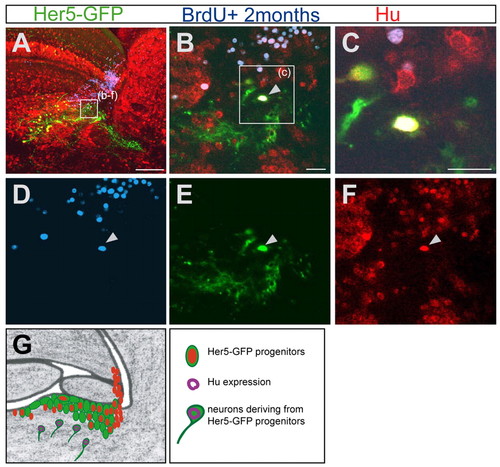
Her5-GFP-positive newborn tegmental neurons originate from cells proliferating at adulthood. (A-F) Sagittal section (anterior is to the left) of a 5-month-old brain stained for Her5-GFP (green), Hu (red) and BrdU (blue), 2 months after a cumulative BrdU labelling. (A) Overview of the tegmental region containing differentiated Her5-GFP-positive cells. (B-F) Higher magnifications of the same section. The arrow in B points to a cell in the tegmentum triply labelled for Hu, BrdU and GFP, indicating that this neuron is deriving from a Her5-expressing cell and was generated 2 months earlier. The single channels of this confocal plane are depicted in D-F. (C) Higher magnification of the same triple-labelled cell. (G) Summary scheme of the IPZ area (as in Fig. 1J, Fig. 2H, Fig. 3F), depicting the tegmental neurons (Hu staining, purple) generated by Her5-expressing progenitor cells. Scale bars: 100 μm in A; 10 μm in B,C. |