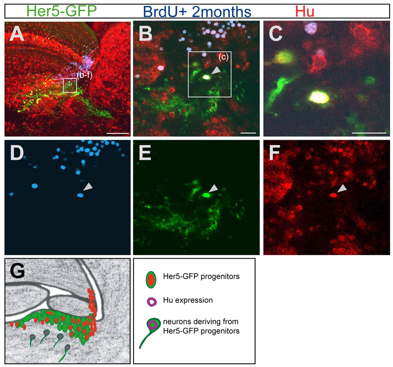
Fig. 7 Her5-GFP-positive newborn tegmental neurons originate from cells proliferating at adulthood. (A-F) Sagittal section (anterior is to the left) of a 5-month-old brain stained for Her5-GFP (green), Hu (red) and BrdU (blue), 2 months after a cumulative BrdU labelling. (A) Overview of the tegmental region containing differentiated Her5-GFP-positive cells. (B-F) Higher magnifications of the same section. The arrow in B points to a cell in the tegmentum triply labelled for Hu, BrdU and GFP, indicating that this neuron is deriving from a Her5-expressing cell and was generated 2 months earlier. The single channels of this confocal plane are depicted in D-F. (C) Higher magnification of the same triple-labelled cell. (G) Summary scheme of the IPZ area (as in Fig. 1J, Fig. 2H, Fig. 3F), depicting the tegmental neurons (Hu staining, purple) generated by Her5-expressing progenitor cells. Scale bars: 100 μm in A; 10 μm in B,C.
Image
Figure Caption
Acknowledgments
This image is the copyrighted work of the attributed author or publisher, and
ZFIN has permission only to display this image to its users.
Additional permissions should be obtained from the applicable author or publisher of the image.
Full text @ Development

