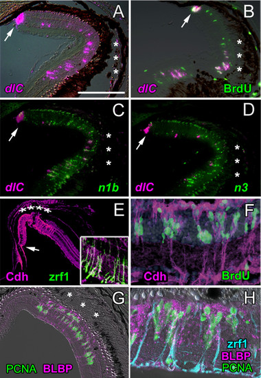
Molecular profile of the injury-induced proliferating retinal progenitors is similar to retinal stem cells in the CMZ. A) At 4 dpl deltaC expression (dlc; magenta) is upregulated in the CMZ (arrow) and in cells in the INL beneath the lesion (asterisks). Scale bar = 150 μm. B) At 4 dpl, cells in the CMZ (arrow) and lesioned area labeled with a 2-hour pulse of BrdU (green) also express deltaC (magenta). C) notch1b (n1b; green) and D) notch3 (n3; green) are also up-regulated but are generally not co-expressed with deltaC (magenta). E) At 4 dpl, N-cadherin immunoreactivity (Cdh2; magenta) is strongly up-regulated in the lesioned area (asterisks) and diffusely localized. Inset: zrf1, anti-zebrafish-GFAP (green) in radial fibers of Müller glia in the lesioned retina (7 dpl) co-localizes with N-cadherin immunoreactivity (magenta). F) At 7 dpl, BrdU+ nuclei (green) associate with Müller glial radial fibers that are strongly immunoreactive for N-cadherin (magenta). DAPI (blue). G) At 3 dpl, activated Müller glia confined to the lesioned region (asterisks) express BLBP (magenta), a marker of immature Müller glia, and they are mitotically active (PCNA+, green). H) Radial fibers of injury-activated Müller glia are zrf1+ (blue). Proliferating Müller nuclei are PCNA+ (green) and many have migrated to the apical surface (the former ONL where photoreceptors are missing) and are BLBP+ (magenta).
|
