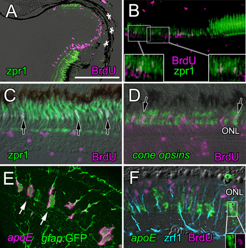
Injury-induced retinal progenitors regenerate cone photoreceptors within a week. A) At 4 dpl, proliferating cells (BrdU; magenta) fill the lesioned area (asterisks) in which double cones immunoreactive for zpr1 (green) are missing. Scale bar = 250 μm. B) By 7 dpl, some retinal progenitors that were labeled with BrdU (magenta) at 4 dpl have begun to differentiate into cones and are double-labeled with zpr1 (green). Boxed areas are shown at higher magnification in the insets; double-labeled cells are white. C) By 31 dpl fully differentiated, regenerated cone photoreceptors (zpr1; green) are labeled with BrdU (magenta) injected at 3 dpl (double-labeled white nuclei are indicated by black arrows). Unidentified BrdU+ nuclei are seen in the inner retina. D) Cocktail of riboprobes to cone opsins (green) identifies BrdU+ (magenta), regenerated cones at 31 dpl (double-labeled white nuclei are indicated by black arrows). BrdU+ rod nuclei (magenta) are in the inner part of the outer nuclear layer, ONL. E) Müller glia in a transgenic zebrafish Tg(gfap:GFP)mi2001 are labeled with anti-GFP (green) and co-express apoE (magenta, in situ hybridization) in their cell bodies. F) At 4 dpl, most BrdU+ proliferating nuclei are in the outer nuclear layer, ONL, but a few apoE+ Müller glial cells are also BrdU+ (inset). Radial fibers of Müller glia are labeled with zpr1/anti-GFAP (blue).
|
