
Fig. 6
- ID
- ZDB-FIG-060824-6
- Publication
- Gulati-Leekha et al., 2006 - A reporter-assisted mutagenesis screen using α1-tubulin-GFP transgenic zebrafish uncovers missteps during neuronal development and axonogenesis
- Other Figures
- All Figure Page
- Back to All Figure Page
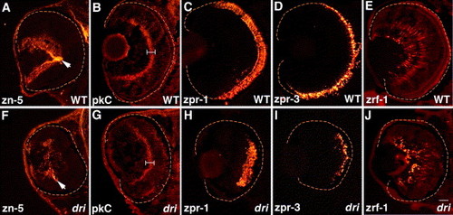
drishti does not affect retinal cell type specification, migration, or terminal differentiation. Retinal sections immunostained with zn-5 (RGCs and optic nerve (arrow) at 3 dpf; A, F); protein kinase C (pkC, bipolar cells and their processes at 4 dpf; (B, G) brackets indicate the strongly staining inner plexiform layer); Fret43 (zpr-1) (double-cone photoreceptors at 5 dpf; C, H); Fret11 (zpr-3) (rod photoreceptors at 5 dpf; D, I); and zrf-1 (Muller glia at 5 dpf; E, J). All cell types examined are present (in reduced numbers) in dri retinas at their expected radial positions and have normal cellular morphologies. Scale bar: 25 μm in panel J for all images. |
| Gene: | |
|---|---|
| Fish: | |
| Anatomical Terms: | |
| Stage: | Protruding-mouth |
Reprinted from Developmental Biology, 296(1), Gulati-Leekha, A., and Goldman, D., A reporter-assisted mutagenesis screen using α1-tubulin-GFP transgenic zebrafish uncovers missteps during neuronal development and axonogenesis, 29-47, Copyright (2006) with permission from Elsevier. Full text @ Dev. Biol.