
Fig. 5
- ID
- ZDB-FIG-060824-5
- Publication
- Gulati-Leekha et al., 2006 - A reporter-assisted mutagenesis screen using α1-tubulin-GFP transgenic zebrafish uncovers missteps during neuronal development and axonogenesis
- Other Figures
- All Figure Page
- Back to All Figure Page
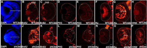
The dri retina is enriched for proliferating retinoblasts, which when unable to withdraw from the cell cycle, undergo premature death. DAPI-stained retinal sections at 3 dpf illustrate the loss of lamination in dri mutants (A, I). Bracketed regions mark the three cellular and two plexiform layers in wild-type retinas replaced by a contiguous neuroepithelial structure. Panels B and J show the localization of BrdU-positive cells in the CGZ of wild-type retinas at 2 dpf whereas a majority of cells in mutant retina are cycling. Albeit restricted to retinal periphery, an expanded S-phase cycling population is still maintained in mutant retinas at 3 dpf (C, K). Arrowheads indicate an accompanying abundance of PH3-positive mitotic figures at 2 dpf (D, L) and 3 dpf (E, M). Developmental progression of retinoblasts to Hu-positive postmitotic cells is severely impaired at 2 dpf, partial recovery observed at 4 dpf with signs of primitive lamination (F, N; G, O, brackets). Excess cells are eliminated by apoptosis in 2-day-old dri retinas illustrated by cleaved-caspase-3 (casp) immunostaining (H, P). Cell death is very rarely detected at later time points examined. Scale bars: 25 μm in all panels. |
| Genes: | |
|---|---|
| Fish: | |
| Anatomical Term: | |
| Stage Range: | Long-pec to Day 4 |
Reprinted from Developmental Biology, 296(1), Gulati-Leekha, A., and Goldman, D., A reporter-assisted mutagenesis screen using α1-tubulin-GFP transgenic zebrafish uncovers missteps during neuronal development and axonogenesis, 29-47, Copyright (2006) with permission from Elsevier. Full text @ Dev. Biol.