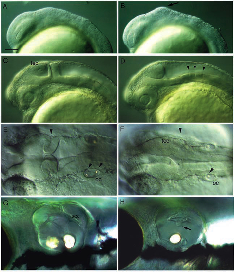
Development of living ace embryos. Shown are lateral or dorsal (E,F) views of whole-mounted living wild type (left) and ace mutant siblings (right) as indicated. (A,B) At the 5-somite stage, mutant embryos show a bulge in the prospective midbrain primordium (arrow). (C,D) At the pharyngula stage, mutant embryos lack a cerebellum and the MHB fold, but show an enlarged tectum. The embryos were placed into PTU to suppress melanization. Arrowheads point to rhombomere boundaries. (E,F) In a dorsal view at 30 h, the absence of the MHB fold and the increased size of the tectum are particularly evident. Arrowheads point to the posterior edge of the tectum, coincident with the MHB fold in the wild type, and to the otoliths in the otic cyst. Notice the reduced size of the otic cyst in the mutant. (G,H) High magnification view of the otic capsule of the free swimming larva (day 6): notice the smaller size, and the truncated semicircular canal (arrow in H). Only one otolith is apparent in the mutant embryo. cb, cerebellum; oc, otic cyst; scc: semicircular canal; tec, tectum. Scale bar, 100 µm (A-D); 75 µm (E,F); 65 µm (G,H).
|
