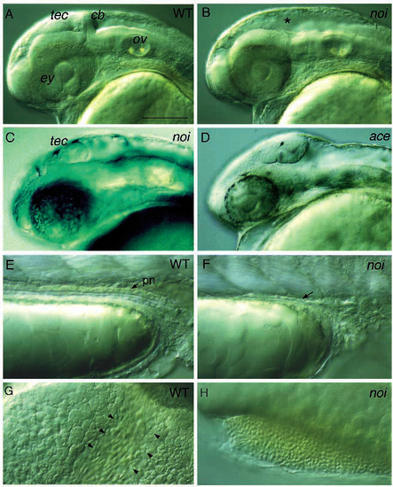
Lateral views of living noi and ace mutant embryos. (A) Wild-type embryo at 36 hours of development. (B) Sibling of the embryo in A that is homozygous mutant for a strong allele of noi (noith44). Absence of the tectum, cerebellum and MHB is indicated by an asterisk. (C) Embryo homozygous mutant for a weak allele of noi (noity31a). A partially formed tectum, but no cerebellum or MHB, is observed. (D) Embryo homozygous mutant for acerebellar (aceti282a). An enlarged tectum is present, but no cerebellum or MHB are seen (see also Fig. 7). (E) Pronephric duct (pn) in a wild type and (F) homozygous mutant embryo for noith44. In the mutant embryos, no pronephric duct is found; the arrow points to the few cells in the mutant that may represent partially developed pronephric duct cells; consistent with the presence of a normal duct during earlier stages of development. (G) Blood streaming through the common cardinal vein over the yolk in a 36 h wild-type embryo (bracketed by arrows). (H) Blood cells accumulate in a dent on the ventral yolk sac in a homozygous mutant embryo for noith44. No such accumulation is seen in wild-type embryos. Embryos in A and B and E-F were placed in PTU at about 26 h to suppress formation of melanin. cb, cerebellum; ey, eye; tec, tectum; ov, otic vesicle; pn, pronephric duct. Scale bar, 150 µm (A-D), 60 µm (E-H).
|
