
Fig. 1
- ID
- ZDB-FIG-060717-19
- Publication
- Hollway et al., 2006 - Scube2 mediates Hedgehog signalling in the zebrafish embryo
- Other Figures
- All Figure Page
- Back to All Figure Page
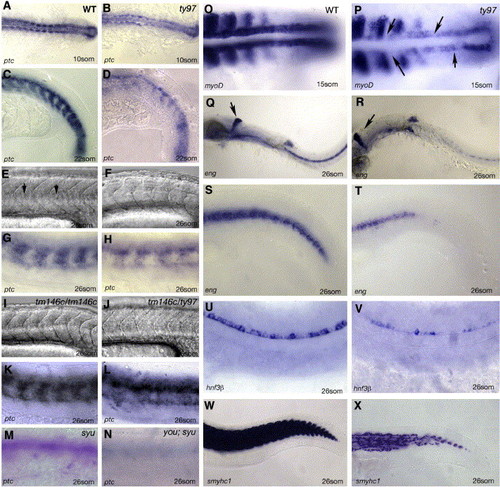
Homozygous you mutants lack HH sensitive cell fates. Panels A–D youty97 mutants show a reduction in ptc expression within the somites and neural tube. Wild type (WT) embryos at the 10-somite (A) or 22-somite stages (C) show robust expression of ptc compared to similar stage homozygous youty97 mutant embryos panels B and D. A–B dorsal views, panels C, D lateral views, anterior to the left. Panels E–L comparative phenotype of different you alleles and intra allelic compounds at 26 hpf. Lateral views anterior to the left. (E) Wildtype myotomes demonstrate a distinct chevron shape and the presence of a horizontal myosepta (arrows). (F) youty97 homozygotes exhibit u-shaped somites and lack a horizontal myosepta. (G) ptc is strongly expressed in wildtype myotomes at this stage (26 hpf). (H) ptc expression is reduced in homozygous youty97 mutants. (I) Homozygous youtm146c mutants exhibit a much less severe alteration in myotomal shape. (J) Intra allelic homozygote compounds between youty97 and youtm146c exhibit a myotomal phenotype intermediate between the homozygote phenotype of the two separate alleles. (K) Myotomal ptc expression is indistinguishable from wild type in homozygous youtm146c mutant embryos. (L) ptc expression in youtm146c/ty97 allelic compound homozygote embryos is reduced compared to youtm146chomozygotes. (M, N) youty97/syudel double mutant embryos exhibit an additive phenotype. (M) 26 somite syudel homozygous mutant embryos exhibit a more severe reduction in somitic ptc staining than homozygous youty97 mutants. (N) Ptc staining is reduced to barely detectable levels within similarly staged double mutant youty97 and syudel embryos. (O, P) myoD expression at the 15-somite stage, specifically within the adaxial cells, is down regulated compared to wildtype (O) in youty97 homozygote embryos (P), arrows indicate gaps in adaxial expression. Panels Q–T engrailed mRNA expression in the pioneer cells at the 26-somite stage is severely down regulated as compared to wildtype (Q, S) within homozygous youty97 mutants (R, T). Arrowheads in panels Q and R mark the midbrain–hindbrain expression of eng which is unaffected within you homozygote mutant embryos. (U, V) Floor plate expression of axial (hnf3β) is reduced or absent compared to wildtype (U) within youty97 homozygote mutant embryos (V). (W, X) At 26 somites, slow myosin heavy chain gene expression is severely reduced compared to wild type embryos (W) within youty97 homozygous mutants embryos (X). |
| Genes: | |
|---|---|
| Fish: | |
| Anatomical Terms: | |
| Stage Range: | 10-13 somites to 26+ somites |
Reprinted from Developmental Biology, 294(1), Hollway, G.E., Maule, J., Gautier, P., Evans, T.M., Keenan, D.G., Lohs, C., Fischer, D., Wicking, C., and Currie, P.D., Scube2 mediates Hedgehog signalling in the zebrafish embryo, 104-118, Copyright (2006) with permission from Elsevier. Full text @ Dev. Biol.