
Fig. 6
- ID
- ZDB-FIG-070918-20
- Publication
- Hollway et al., 2006 - Scube2 mediates Hedgehog signalling in the zebrafish embryo
- Other Figures
- All Figure Page
- Back to All Figure Page
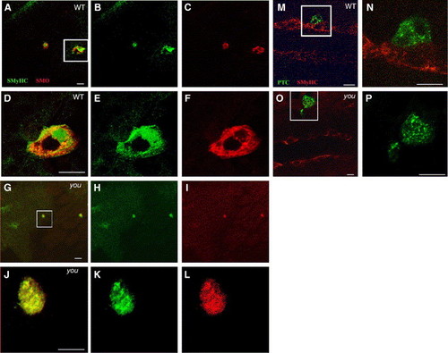
Scube2 is not required for the cytoplasmic localisation of Patched or Smoothened. (A–F) Constitutively active Smoh mutant M1 induces Slow MyHC expression cell autonomously within the PSM of wild type and youty97 mutant embryos and is localised predominately intracellularly within a 15-somite stage embryo. Oblique, dorsal confocal sections at the level of the notochord anterior to the top. (A) Slow MyHC (SMyHC) expression (green) within the adaxial cells and ectopically within activated Smoh M1 (red) clones. (B) SMyHC alone. (C) Smoh protein expression alone. (D–F) High magnification views of the single cell expressing activated Smoh boxed in panel A. (D) Slow MyHC (SMyHC) expression (green) within the activated Smoh M1 (red) clones, revealing a predominately intracellular localisation within induced clones. (E) SMyHc alone. (F) Smoh protein expression alone. (G–L) Induction of slow MyHC expression and Smoh localisation are unaffected when mosaic Smoh M1 clones are induced within a 15-somite stage homozygous youty97 mutant embryo. (G) Mosaic Smoh M1 expression (red) induces slow MyHC expression (green) in a youty97 homozygote embryo. (H) SMyHc alone. (I) Smoh protein expression alone. (J–L) High magnification views of the single cell, boxed in panel G, expressing activated Smoh and activated Smoh M1 (red). (J) Slow MyHC (SMyHC) expression (green) within the activated Smoh M1 (red) clones. (K) SMyHc alone. (L) Smoh protein expression alone, revealing a predominately intracellular localisation of Smoh protein within expressing clones, similar to clones induced in wildtype embryos. (M, N) Ptc distribution within a wildtype 15-somite stage embryo, monitored using C-terminally fused GFP is constitutively intracellular, regardless of localisation of Ptc expressing clones within the PSM. Dorsal confocal sections at the level of the notochord, anterior to the left. (M) SMyHC (red) denotes the position of the adaxial cells with a single Ptc (Green) expressing cell localised immediately lateral to the adaxial cells. (N) High magnification view of the region boxed in panel M revealing Ptc is localised primarily to intracellular vesicular structures suggestive of endosomal elements, consistent with previous observations (Incardona et al., 2002 and Evans et al., 2003). (O, P) Localisation of PTC within intracellular foci does is not altered by expressing PTC-GFP within a youty97 homozygous mutant embryo. (P) High magnification view of the region boxed in panel O. Panels O and P are similar stages and views to those shown in panels M, N. Scale bars, 20 μm. |
Reprinted from Developmental Biology, 294(1), Hollway, G.E., Maule, J., Gautier, P., Evans, T.M., Keenan, D.G., Lohs, C., Fischer, D., Wicking, C., and Currie, P.D., Scube2 mediates Hedgehog signalling in the zebrafish embryo, 104-118, Copyright (2006) with permission from Elsevier. Full text @ Dev. Biol.