
Fig. 4
- ID
- ZDB-FIG-070918-18
- Publication
- Hollway et al., 2006 - Scube2 mediates Hedgehog signalling in the zebrafish embryo
- Other Figures
- All Figure Page
- Back to All Figure Page
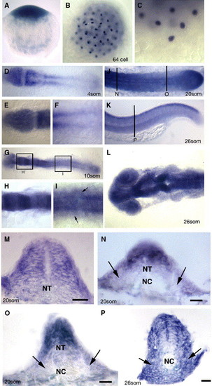
Embryonic expression of zebrafish scube2. (A–P) Expression of the gene (blue) encoded by the you locus as revealed by in situ hybridisation. A. you mRNA is deposited maternally as evidenced by the detection of mRNA in the one cell stage embryo. Lateral view, dorsal to the top. (B) Maternally deposited you mRNA is localised to discrete regions in cleavage stage embryos, as evidenced by in situ hybridisation on this 64 cell stage embryo. Dorsal view. (C) High magnification view of the most ventral cells of the same embryo as in (B). Lateral view dorsal to the top. (D) 4-somite stage embryos show strong expression of you mRNA in the anterior neural plate, newly formed somites and PSM. (E) High magnification of you expression in the neural plate at the same stage as in (D). (F) High magnification at the same stage as in (D) of expression within PSM. (G) Expression of you mRNA at the 10-somite stage reveals a modulation of expression within different regions of the hindbrain (high magnification view in panel H), and continued expression within the neural tube and PSM. Expression within the posterior neural tube is also evident at this stage making expression within the neural epithelium global (High magnification view in (I), at the dorsal ventral-level of the notochord, arrows indicate expression within somitic mesoderm cells). (J) Expression at the 20-somite stage continues in the neural tube, somites and PSM, including robust expression within the tail bud (arrow). (D–J dorsal views anterior to the left). (K) At the end of somitogenesis (26-somite stage), expression within the posterior neural tube and myotome is down regulated with the exception of the dorsal most aspect of the neural tube. Lateral view anterior to the left. Vertical line denotes level of the section (P). (L) At 26 somites, the anterior neural tube expression remains high, and modulates within specific neuronal regions, dorsal view anterior to the left. (M) Transverse section of the anterior neural tube of 20-somite stage embryo revealing expression throughout the entire neural epithelium. Panels N–P transverse section from the locations and stages indicated in panels J and K, Arrows demark the somitic expression of zebrafish scube2. Scale bars 20 μM. PSM—presomitic mesoderm, NT—neural tube, NC—notochord. |
Reprinted from Developmental Biology, 294(1), Hollway, G.E., Maule, J., Gautier, P., Evans, T.M., Keenan, D.G., Lohs, C., Fischer, D., Wicking, C., and Currie, P.D., Scube2 mediates Hedgehog signalling in the zebrafish embryo, 104-118, Copyright (2006) with permission from Elsevier. Full text @ Dev. Biol.