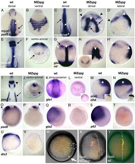
Meso- and ectodermal gene expression in MZspg embryos.(A,C) myod is expressed in wild-type somites at the eight-somite stage (white arrow). (B,D,D') Somitic myod expression is displaced and fuses ventrally in MZspg (black arrowheads in D,D'). (A,B,I,J) Pronephric pax2.1 expression (black arrows in A,I) is missing in MZspg. (C,E white arrows; G white arrowhead) ntl is expressed in the notochord and tb in wild-type embryos. (D,D',H,H', white arrowheads) ntl expression is broadened and variably split in MZspg embryos. Single ntl expressing cells are found in close proximity to the notochord (black arrows in D). (E,F) krox20 expression in rhombomere 3 and 5 (arrow) is severely reduced, and residual expression fuses at the ventral side. (G,H,H') gbx2 expression is absent at the MHB in MZspg. Mesodermal gbx2 expression (white arrows) fuses at the ventral side in MZspg. Inset in H shows ventro-animal view of MZspg solely expressing gbx2, which fuses at the ventral side. (I,J) pax2.1 expression at the MHB (arrowhead) is strongly reduced and fuses at the ventral side. (K,L) gata1 is normally expressed in blood progenitor cells. gata1 is strongly reduced in MZspg. Inset shows a lateral view of the same embryo. (M-V) Lateral views. (M,N) At tb stage otx2 (asterisk) is expressed in the wild-type forebrain and is strongly expanded to the ventral side in MZspg, manifested already at the initial phase of its expression (insets, 40-50% E). (O,P) pax6 is expressed within the wild-type forebrain and hindbrain at tb stage. pax6 domains are radialized in MZspg as indicated by white and black asterisks. (Q-V) MZspg embryos lack gata2, p63 and dlx3 expression at the end of gastrulation. (W-Y) Cell movement behavior. Bright-field images of transplanted living embryos were merged with fluorescent images taken at the same focal plane. Transplanted wild-type and MZspg cells are visualized by red or green fluorescence, respectively. (W) Animal view of a chimeric embryo transplanted at the shield stage carrying wild-type and MZspg cells in the dorsolateral germring (white arrow). A broken circle outlines the shield. (X,Y) Dorsal views of tb stage host embryos, which were transplanted as the embryo depicted in W. Broken lines indicate dorsal AP axes, anterior is towards the top. (X,Y) MZspg mutant cells are indistinguishable in their movement behavior from co-transplanted wild-type cells. (X) In MZspg embryos, convergence towards the dorsal midline and extension along the AP axis is affected in both transplanted MZspg and wild-type cells. (Y) Convergence-extension of transplanted MZspg and wild-type cells is normal in wild-type embryos.
|
