|
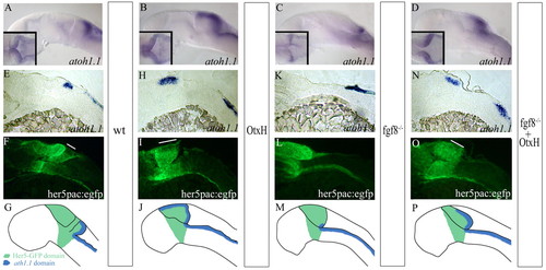
Formation of the granule cell population in fgf8-/-; OtxH embryos. atoh1a expression in wild-type (A), OtxH (B), fgf8-/- (C) and fgf8-/-; OtxH (D) whole-mount embryos. MyoD expression in the somite was concomitantly detected to confirm fgf8-/-genotype. (E-O) Para-saggital 10 μm sections, anterior towards the left, of wild-type her5pac:egfp (E,F), OtxH (H,I), fgf8-/- (K,L) and fgf8-/-; OtxH (N,O) brains, showing co-localisation of atoh1a (E,H,K,N) and GFP expression (F,I,L,O). Granule cells absent in fgf8-/- are rescued in fgf8-/-; OtxH and are GFP positive (white lines on fluorescent pictures represent the length of the I>atoh1a domain in the upper rhombic lip obtained from bright field pictures). (G,J,M,P) Cartoons summarising GFP (in green) and atoh1a (in blue) expressions in wild-type (G), OtxH (J), fgf8-/- (M) and fgf8-/-; OtxH (P) embryos carrying the transgene her5pac:egfp.
|