
Fig. 6
- ID
- ZDB-FIG-051104-9
- Publication
- Picker et al., 2005 - Fgf signals from a novel signaling center determine axial patterning of the prospective neural retina
- Other Figures
- All Figure Page
- Back to All Figure Page
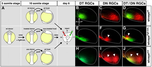
Topographic shifts of tectal RGC projections from eyes with altered Fgf signaling. Retinotectal topography in chimeric larvae after transplantation of optic vesicles with Fgf-dependent alterations in NT patterning. (A) Transplantation scheme: optic vesicles were grafted from mock-treated wild-type donors to wild-type hosts (top), from Fgfr-inhibitor-treated donor embryos (treatment: 5- to 10-somite stage) to non-treated hosts (middle) and from ace donors to wild-type hosts (bottom) at the 10-somite stage. Chimeras were fixed at day 6 and retinal ganglion cell (RGC) axons anterogradely double-labeled by DiI (green) and DiO (red) in the dorsotemporal (DT) and dorsonasal (DN) quadrants. (B-D) In control transplantations, DT RGCs form an anterior termination zone (TZ) and DN RGCs form a posterior TZ that is non-overlapping and correct, according to their NT position in the retina (n=6/6). (E-G) Eyes from Fgfr-inhibitor-treated donor embryos display a full temporalization of RGC axon mapping: DT RGCs correctly terminate in an anterior TZ, while DN RGCs misproject to an ectopic anterior TZ (n=5/5). (H-J) Eyes from ace donor embryos display a partial temporalization of RGC axon mapping: DT RGCs correctly terminate anteriorly, while DN RGCs form a second, ectopic anterior TZ (n=5/6). White arrowheads indicate misprojections compared with wild type. Orientation of the tectum (dorsal views) as indicated in B. (B-J) Maximum-intensity projections of confocal image stacks. Broken outlines indicate the tectal neuropil. A, anterior; L, lateral; M, medial; P, posterior. |