
Fig. 1
- ID
- ZDB-FIG-051104-4
- Publication
- Picker et al., 2005 - Fgf signals from a novel signaling center determine axial patterning of the prospective neural retina
- Other Figures
- All Figure Page
- Back to All Figure Page
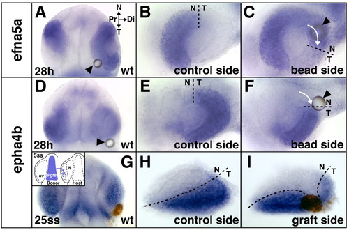
Temporal shifts in retinal patterning induced by ectopic Fgf8. (A-F) Shifts in retinal NT gene expression at the 28 hour stage after Fgf8-bead implantation at the 5-somite stage into wild-type (wt) embryos: expansion of the nasal marker efna5a (A,C) and reduction of the temporal marker epha4b (D,F) compared with control side of the same embryos (B,E). (G-I) Reduction of temporal epha4b expression in a wild-type embryo at the 25-somite stage after ectopic transplantation of cells from the telencephalic Fgf8-domain into the optic vesicle (ov, brown staining) at the 5-somite stage (I) compared with control side of the same embryo (H) (see inset in G for grafting schematic). Arrows indicate directionality of expansion/reduction. Location of the bead is indicated by arrowheads. (A,D,G) Dorsal views with anterior towards the top, orientation as indicated in A; (B,C,E,F,H,I) flat-mounts of dissected eyes. (C,F,I) Digitally inverted images. N, nasal; T, temporal; Pr, proximal; Di, distal. Dashed lines indicate the retinal NT boundary. |