
Fig. 7
- ID
- ZDB-FIG-051104-10
- Publication
- Picker et al., 2005 - Fgf signals from a novel signaling center determine axial patterning of the prospective neural retina
- Other Figures
- All Figure Page
- Back to All Figure Page
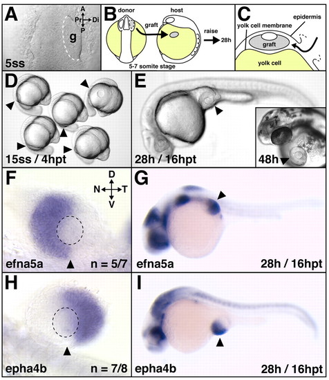
Correct retinal NT patterning after heterotopic optic vesicle transplantation. (A-C) Transplantation scheme. At the 5- to 7-somite stage, optic vesicles were transplanted from the forebrain of donor embryos onto the yolk cell of hosts (B) by slipping the graft in between the locally-opened epidermis and the yolk cell membrane (C). A live image of the anterior neural keel (dorsal view) in shown in A, (D,E) Live host embryos: heterotopic grafts develop and differentiate on the yolk cell and form eyes of normal morphology. (F-I) Retinal NT patterning after heterotopic transplantation at 28 hours of development: in grafts, the nasal expression domain of efna5a (F) and the temporal expression domain of epha4b (H) are established and maintained normally in the ectopic situs (G,I) of the embryo. (F,H) Flat-mounts of dissected eyes, orientation as indicated in F. Arrowheads indicate the implantation site (D,E,G,I) and the ventralmost position of the eye (F,H). Broken outline in A indicates grafted tissue; in F,H it indicates the lens. A, anterior; P, posterior; g, graft; hpt, hours post-transplantation; D, dorsal; V, ventral; N, nasal; T, temporal; Pr, proximal; Di, distal. |