
Fig. 5
- ID
- ZDB-FIG-050729-7
- Publication
- Xiao et al., 2005 - A GFP-based genetic screen reveals mutations that disrupt the architecture of the zebrafish retinotectal projection
- Other Figures
- All Figure Page
- Back to All Figure Page
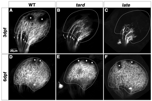
tarde demais (tard) and late bloomer (late) mutants show mildly delayed innervation of the tectum. (A-F) Confocal images of Brn3c:mGFP labeled retinotectal projections of 80 hpf tecta in live larvae (A-C). The wild-type tectum (A) is filled with retinal axons. The optic tract has branched into stereotyped fascicles, labeled with numbers. The tard tectum (B) and the late tectum (C) are less than halfway innervated. (D-F) Lateral views of 6 dpf tecta. The tard tectum and the late tectum are now covered with axons. However, the tard tectum is small and its boundary remains abnormal (E), compared with wild type (D) and in contrast to late (F). Asterisks indicate melanophores on the skin. Broken lines outline the tectal neuropil. Scale bars: 20 µm.
|