
Fig. 8
- ID
- ZDB-FIG-050729-22
- Publication
- Xiao et al., 2005 - A GFP-based genetic screen reveals mutations that disrupt the architecture of the zebrafish retinotectal projection
- Other Figures
- All Figure Page
- Back to All Figure Page
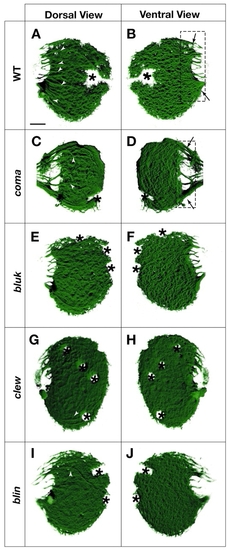
Fasciculation is disorganized in blue kite (bluk) and coming apart (coma), blue kite (bluk), clewless (clew) and blind date (blin) mutants. (A-F) Surface-rendered 3D reconstructions of confocal images taken from 6 dpf live Brn3c:mGFP larvae. Dorsal views (A,C,E,G,I) and mirror-image ventral views (B,D,F,H,J) of the same tecta are shown. On the dorsal surface, axon fascicles are visible within the tectal neuropil of wild type (A) (arrowheads). The ventral view (B) demonstrates the characteristic dense grid of arbors. (C,D) In coma, fascicles both in the optic tract (hatched rectangle in D) and in the neuropil are disorganized. (E-J) In bluk, clew and blin, fascicles within the neuropil are greatly reduced (E-I) and axon termations appear diffuse. Asterisks indicate melanophores in the skin. Scale bars: 20 µm.
|