
Fig. 3
- ID
- ZDB-FIG-050214-9
- Publication
- Galloway et al., 2005 - Loss of gata1 but not gata2 converts erythropoiesis to myelopoiesis in zebrafish embryos
- Other Figures
- All Figure Page
- Back to All Figure Page
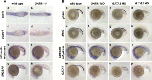
Erythroid Genes Are Differentially Regulated by Gata1 and Gata2(A) Embryos (a–d) have been flat-mounted and photographed to show only the posterior tail region (anterior, left; posterior, right). Normal expression of gdpbp1 and epsin (a, c) is lost in ICM erythroid precursors of vlt mutants at 18 hpf (b, d). Expression of biliverdin reductase and SH3BP5 at 20 hpf (e) and 22 hpf (g), respectively, is absent in vlt mutant ICM cells (f, h).(B) In situ hybridization for βe1 globin (a–d), alas2 (e–h), carbonic anhydrase (i–l), and gata1 (m–p) at 20 hpf. Compared to wild-type embryos (a, e, i, m), ICM expression of βe1 globin, alas2, carbonic anhydrase, and gata1 was decreased in gata1 morphants (b, f, j, n) and absent in g1/g2 morphants (d, h, l, p). Decreased number of cells expressing βe1 globin, alas2, carbonic anhydrase, and gata1 are observed in gata2 morphants (c, g, k, o) compared to wild-types (a, e, i, m). |
| Genes: | |
|---|---|
| Fish: | |
| Knockdown Reagents: | |
| Anatomical Terms: | |
| Stage Range: | 14-19 somites to 26+ somites |
Reprinted from Developmental Cell, 8(1), Galloway, J.L., Wingert, R.A., Thisse, C., Thisse, B., and Zon, L.I., Loss of gata1 but not gata2 converts erythropoiesis to myelopoiesis in zebrafish embryos, 109-116, Copyright (2005) with permission from Elsevier. Full text @ Dev. Cell