
Fig. S2
- ID
- ZDB-FIG-080730-34
- Publication
- Galloway et al., 2005 - Loss of gata1 but not gata2 converts erythropoiesis to myelopoiesis in zebrafish embryos
- Other Figures
- All Figure Page
- Back to All Figure Page
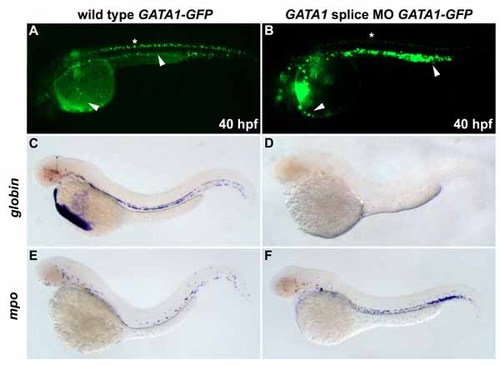
Wild-type gata1-egfp embryos have GFP in circulating blood cells in the vessels and over the yolk (A, arrowheads) and ectopically in the neural tube (asterisk). A gata1 morpholino targeting the exon/intron splice sites was used to avoid interference with transgene expression. At 48 hpf, Gata1-deficicent embryos had large, round, noncirculating GFP-positive cells distributed in a pattern resembling mpo expression (B). gata1 MO-injected gata1-egfp embryos express gfp in noncirculating cells in the major vessels and on the yolk (B, arrowheads). be1 globin (C) and mpo (E) are expressed normally in wild-type embryos. In gata1 MO-injected embryos, be1 globin expression is absent (D) and mpo expression is expanded (F). |
Reprinted from Developmental Cell, 8(1), Galloway, J.L., Wingert, R.A., Thisse, C., Thisse, B., and Zon, L.I., Loss of gata1 but not gata2 converts erythropoiesis to myelopoiesis in zebrafish embryos, 109-116, Copyright (2005) with permission from Elsevier. Full text @ Dev. Cell