
- Title
-
The zebrafish detour gene is essential for cranial but not spinal motor neuron induction
- Authors
- Chandrasekhar, A., Schauerte, H.E., Haffter, P., and Kuwada, J.Y.
- Source
- Full text @ Development
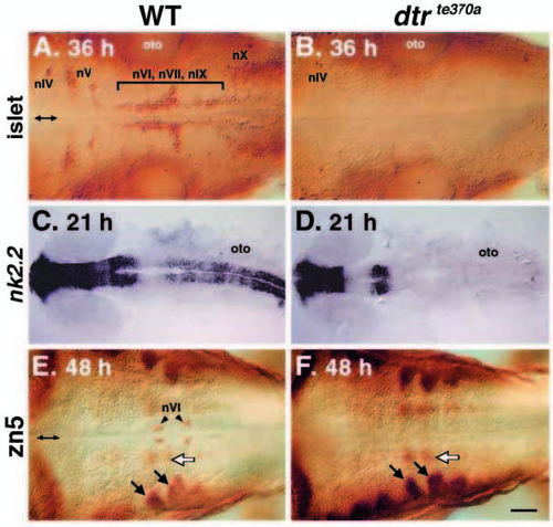
Cranial motor neurons are missing in dtrte370a embryos. All panels depict dorsal views, with rostral to the left, of the hindbrain of whole-mounted embryos analyzed either by islet (A,B) or zn5 (E,F) immunohistochemistry, or by nk2.2 in situ hybridization (C,D). Double arrows (A,E) mark the midline. (A) In a 36 HPF wild-type sibling, the islet antibody labels the trigeminal motor (nV) neurons in r2 and r3, the abducens (nVI), the facial motor (nVII) and the glossopharyngeal motor (nIX) neurons in r4, r5, r6 and r7, and the vagal motor (nX) neurons in the caudal hindbrain. The antibody also labels the presumptive trochlear (nIV) neurons in r1. (B) In a dtrte370a homozygote, all cranial motor neurons, except the putative nIV neurons, are missing. (C) In a 21 HPF wild-type sibling, nk2.2 is expressed in the ventral CNS throughout the forebrain, the rostral midbrain and the hindbrain. (D) In a dtrte370a homozygote, nk2.2 expression is missing throughout the hindbrain and in the rostralmost midbrain. (E) In a 48 HPF wild-type sibling, the zn5 antibody labels the abducens neurons (nVI) in r5 and r6, and some unidentified cells just laterally (white arrow). The labeled cells located most laterally (black arrows) are the hindbrain commissural neurons. (F) In a dtrte370a homozygote, the nVI neurons are missing. However, the hindbrain commissural neurons (black arrows) and the unidentified zn5-labeled cells (white arrow) are unaffected. oto, otocyst. Scale bar, 40 μm (A,B,E,F), 100 μm (C,D). EXPRESSION / LABELING:
|
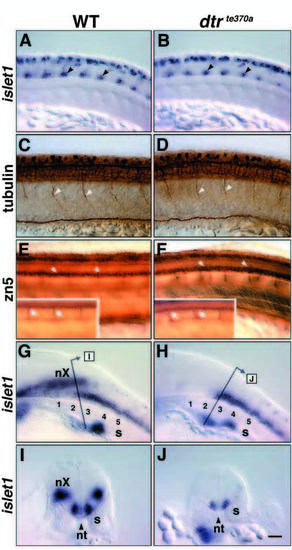
Spinal motor neurons are generated normally in dtrte370a embryos. Panels A-H depict lateral views, with rostral to the left and dorsal up, of the trunk of whole-mounted embryos analyzed either by anti-tubulin (C,D) or zn5 (E,F) immunohistochemistry, or by islet1 in situ hybridization (A,B,G,H). The right-angled arrows in G and H indicate the approximate location and orientation of the transverse sections shown in I and J, respectively, obtained from different embryos. (A) In a 21 HPF wild-type sibling, the two to three isl1- expressing cells (arrowheads) in the ventral spinal cord in every hemisegment are the primary motor neurons. The isl1-expressing cells in the dorsal spinal cord are the Rohon-Beard neurons. (B) In a dtrte370a homozygote, the primary motor neurons (arrowheads) and Rohon-Beard neurons appear normal in number and location. (C) In a 24 HPF wild-type sibling, the primary motor axons exit the spinal cord, with one motor root per hemisegment (arrowheads). (D) In a dtrte370a homozygote, the number and appearance of the primary motor axons exiting the spinal cord (arrowheads) are unaffected. (E) In a 48 HPF wild-type sibling, the zn5-labeled secondary motor neurons are located in the ventral fourth of the spinal cord (arrowheads). Inset depicts a more lateral focal plane showing the secondary motor axons (arrows) exiting the spinal cord and extending ventrally into the somites. (F) In a dtrte370a homozygote, the secondary motor neurons (arrowheads) appear normal in number. However, many secondary motor axons (Inset, arrows) exit the spinal cord at ectopic locations. (G) In a 30 HPF wild-type sibling, the caudalmost nX neurons overlap the rostralmost spinal motor neurons located at the level of somites 2 and 3. (H) In a dtrte370a homozygote, the nX neurons are missing, but the rostralmost spinal motor neurons are still present. (I) Transverse section through the caudal hindbrain in a wild-type sibling shows that the nX neurons and the spinal motor neurons overlap rostrocaudally, but occupy distinct dorsolateral locations. (J) Transverse section through the caudal hindbrain in a dtrte370a homozygote reveals only the rostralmost spinal motor neurons. The apparent difference in isl1 expression in spinal motor neurons between I and J results from the different thicknesses of the sections, which were done by hand. s, somite; nt, notochord. Scale bar, 40 μm. EXPRESSION / LABELING:
|
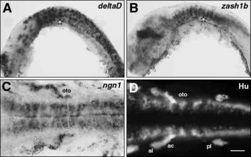
Neurogenesis occurs normally in dtrte370a mutants. (A,B) Lateral views, and (C,D) dorsal views, with rostral to the left, of whole-mounted embryos analyzed either by in situ hybridization (A-C) or by immunohistochemistry (D). The embryos used in each experiment were obtained from a dtrte370a/+ incross, and all embryos exhibited similar labeling indicating that these markers were expressed in a similar fashion in wild-type and mutant embryos. A representative embryo is shown in each case. The asterisk (A,B) indicates the location of the otocyst. (A) At 18 HPF, when branchiomotor neurons are beginning to differentiate, deltaD is expressed extensively in the hindbrain at all dorsoventral levels. (B) At 24 HPF, zash1b is expressed strongly in the hindbrain, especially in dorsoventral columns of cells. (C) At 24 HPF, when branchiomotor neurons are normally still being generated, neurogenin1 is expressed in a broad longitudinal column of cells located medially in the ventral hindbrain. (D) At 24 HPF, the Hu antibody labels cells within longitudinal columns located laterally within the ventral hindbrain. ac, acoustic ganglion; al, anterior lateral line ganglion; oto, otocyst; pl, posterior lateral line ganglion. Scale bar, (A,B) 100 μm, (C,D) 50 μm. EXPRESSION / LABELING:
|
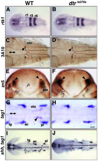
Hindbrain patterning and differentiation of many neurons are normal in dtrte370a mutants. All panels depict dorsal views (except E, F), with rostral to the left, of whole-mounted embryos analyzed either by immunohistochemistry (C-F) or by in situ hybridization (A,B,G-J). Double arrows (C,G) mark the midline. (A) In a 28 HPF wild-type sibling, rtk1 (EphA4) is expressed in r1, r3 and r5. (B) In a dtrte370a homozygote, rtk1 expression is normal. (C) In a 36 HPF wild-type sibling, the 3A10 antibody labels the Mauthner cells (arrowhead) and their axons, which cross the midline and extend caudally into the spinal cord. (D) In a dtrte370a homozygote, the Mauthner cells (arrowhead) and their axons are unaffected. (E) In a 36 HPF wild-type sibling, a transverse section at the level of rhombomere 5 reveals zn5 antibody-labeled commissural neurons (black arrowheads) and their axons (arrow), and the abducens motor neurons medially (white arrowhead). (F) In a dtrte370a homozygote, the commissural neurons (arrowheads) and their axons (arrow) are unaffected, but the abducens motor neurons are missing. The difference in staining intensity between E and F results from the different thicknesses of the sections, which were done by hand. The break in the commissural axons in F is due to a crack in the tissue. (G) In a 20 HPF wild-type sibling, the tag1-expressing presumptive nVII neurons (arrowhead) span r5, r6 and r7. The prominent patches of labeling located laterally, rostral and caudal to the otocyst (oto), represent tag1 expression in cranial sensory ganglia. (H) In a dtrte370a homozygote, the presumptive nVII neurons are missing, but the cranial ganglia are unaffected. (I) In a 24 HPF wild-type sibling, shh is expressed in the floor plate (arrow) throughout the midbrain and hindbrain, and in the ventral neuroectoderm in the forebrain. tag1 expression in the nVII neurons (arrowhead) and in the cranial ganglia is also evident. (J) In a dtrte370a homozygote, the nVII neurons are missing. However, shh is expressed normally in the floor plate (arrow), and the cranial ganglia are unaffected. ac, acoustic ganglion; oto, otocyst; tg, trigeminal ganglion; pl, posterior lateral line ganglion. Scale bar, (A,B,I,J) 100 μm, (C,D) 50 μm, (E,F) 25 μm, (G,H) 40 μm. EXPRESSION / LABELING:
PHENOTYPE:
|
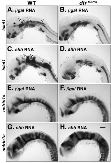
shh overexpression does not induce branchiomotor neurons in dtrte370a embryos. All panels show lateral views of the head, with rostral to the left and dorsal up, in embryos analyzed by islet1 (isl1; A-D) or netrin1a (net1a; E-H) in situ hybridization. Embryos at the 1- to 4-cell stage were injected with full-length lacZ (A,B,E,F) or shh (C,D,G,H) RNA, and fixed at 30 HPF. (A) In a lacZ-injected wildtype sibling, the clusters of nV, nVII and nX neurons (arrows) are evident. The asterisk denotes putative oculomotor (nIII) neurons in the midbrain. (B) In a lacZ-injected dtrte370a homozygote, the branchiomotor, as well as the putative oculomotor, neurons are missing. (C) In a shh-injected wild-type sibling, a large number of ectopic isl1-expressing cells are induced throughout the hindbrain (arrows), which have been shown to be branchiomotor neurons (Chandrasekhar et al., 1998). (D) In a shh-injected dtrte370a homozygote, no isl1-expressing cells are induced in the hindbrain. (E) In a lacZ-injected wild-type sibling, net1a is expressed in the ventral half of the CNS throughout the embryo and in dorsoventral columns at rhombomere boundaries. (F) In a lacZ-injected dtrte370a homozygote, net1a expression is normal. (G) In a shh-injected wildtype sibling, net1a expression in the CNS is expanded dorsally at all rostrocaudal levels. Supernumerary muscle pioneer cells expressing net1a are also evident in the somites, as described previously. (H) In a shh-injected dtrte370a homozygote, net1a is ectopically expressed in the CNS and in the somites, in a similar fashion to injected wild-type embryos. Scale bar, 100 μm. |
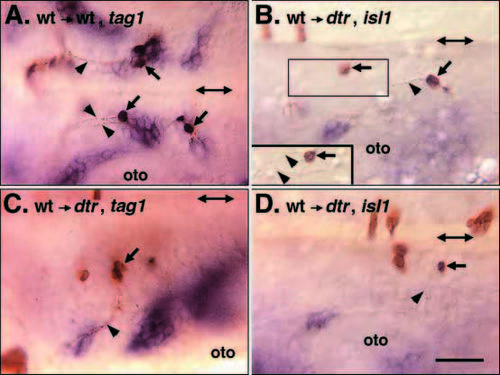
detour functions cell autonomously in branchiomotor neuron induction. All panels show dorsal views of the hindbrain, with rostral to the left, in whole-mounted embryos processed for tag1 (A,C) or islet1 (B,D) in situ hybridization. Double arrows in each panel mark the midline. Donor wildtype cells and their axons are labeled brown. Embryos in B-D were identified as mutant host embryos because their hindbrains did not contain the characteristic clusters of branchiomotor neurons expressing tag1 (C) or islet1 (B,D). (A) When wild-type cells are transplanted into a wild-type host embryo, the donor cells within the nVII neuronal clusters (arrows) extend axons (arrowheads) anteriorly that exit the hindbrain in r4 into the hyoid arch, as described previously (Chandrasekhar et al., 1997). (B) In a dtr host embryo, donor wild-type cells (arrows) in r4 and r6 extend axons (arrowhead) anteriorly. The boxed area is depicted in a different focal plane (inset) to show that the axons (arrowheads) turn laterally exiting the hindbrain in r4 into the hyoid arch, in a manner characteristic of nVII axons. (C) In another dtr host embryo, donor wild-type cells (arrow) in r2 extend axons (arrowhead) laterally that exit the hindbrain in r2 into the mandibular arch, in a manner characteristic of nV axons. (D) In a third dtr host embryo, a donor wild-type cell (arrow) in r6 extends an axon (arrowhead) laterally that exits the hindbrain in r6, in a manner characteristic of nIX axons. oto, otocyst. Scale bar, 40 μm. |