FIGURE SUMMARY
Title

Microinjection of angiotensin II into zebrafish embryos induces transient dilation and elastin disruption of the dorsal aorta

Authors
Tanifuji, S., Uchida, K., Kawahara, G., Nakamura, T., Iida, S., Hayashi, Y.K., Yokoyama, U.
Source
Full text @ Physiol. Rep.

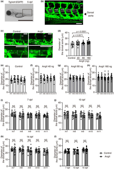
Angiotensin II (AngII) administration increased the dorsal aorta diameter in zebrafish embryos. (a) A lateral bright field image of Tg(kdrl:EGFP) control embryos at 5 days post‐fertilization (dpf). Scale bar: 1 mm. (b) A confocal microscopy image of the EGFP‐positive blood vessels of the boxed region in (a); m7 to m11 indicates myotomes 7–11, respectively. Scale bar: 100 μm. (c) Representative lateral dorsal aorta images of control or AngII (160 ng)‐injected Tg(kdrl:EGFP) embryos at 5 dpf. The lower panels are higher‐magnification images of the upper panels. Scale bars: 50 μm. (d) Quantification of the average diameter of the dorsal aorta. (e–h) Dorsal aorta diameter at myotomes 7–11 in control or AngII (40 ng, 80 ng, or 160 ng)‐injected Tg(kdrl:EGFP) embryos at 5 dpf. n = 15 (control), 15 (40 ng), 13 (80 ng), and 18 (160 ng). (i–k) Dorsal aorta diameter at myotomes 7–11 in control or AngII (160 ng)‐injected Tg(kdrl:EGFP) embryos at 7 dpf (i), 10 dpf (j), and 14 dpf (k). (l) Quantification of the average diameter of the five serial segments of the dorsal aorta. n = 14 (control, white), and n = 14 (AngII 160 ng, gray). NS indicates not significant. Dotted lines in (b) and (c) indicate the diameter of the dorsal aortae. DA, dorsal aorta; DLAV, dorsal longitudinal anastomotic vessel; ISV, intersegmental vessel; PCV, posterior cardinal vein.

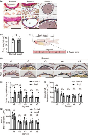
Angiotensin II (AngII) attenuated elastic fiber formation in the adult zebrafish dorsal aorta. (a) Representative images of Elastica van Gieson–stained transverse sections of the dorsal aorta of Tg(kdrl:EGFP) zebrafish at 4, 6, and 8 weeks post‐fertilization (wpf) (The experiment with adult zebrafish included seven zebrafish: Two females and one male at 6 wpf, and two females and two males at 8 wpf. n = 2, 3 (two females and one male), and 4 (two females and two males), respectively). Lower panels indicate higher magnification views of the dorsal aorta in the upper panels. 2 to 3 elastic lamellae were seen at 8 wpf (showed by arrows). DA, dorsal aorta. Scale bars: 20 μm. (b) Body length, length from the tip of the snout to the base of the tail, of control and 160 ng AngII‐injected Tg(kdrl:EGFP) zebrafish at 8 wpf. n = 5 (control) and 8 (160 ng AngII‐injected zebrafish). NS indicates not significant. (c) The illustration indicates the dorsal aorta region, that is, segments v1 through v5, for quantitative analyses. (d) Representative Elastica van Gieson–stained transverse images of the dorsal aorta segments v1 to v5 at 8 wpf. Scale bars: 20 μm. (e–g) Quantification of elastin grading, aorta diameter, and area of the segments v1 to v5 in (d). n = 5 (control) and 8 (160 ng AngII‐injected zebrafish). NS indicates not significant.

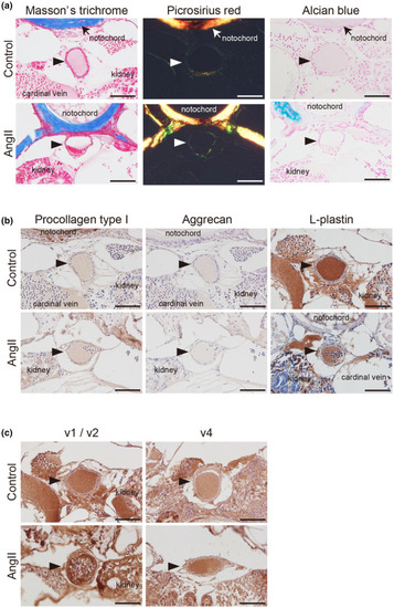
Matrix metalloproteinase (MMP)‐2 upregulation in the dorsal aorta of angiotensin II (AngII)‐injected zebrafish. Representative Masson's trichrome, Picrosirius red, and Alcian blue‐stained (a), immunohistochemically stained with anti‐procollagen type I, aggrecan, and L‐plastin antibodies (b) of the transverse sections taken from the segments v1/v2 of the dorsal aorta in control and 160 ng AngII‐injected Tg(kdrl:EGFP) zebrafish at 8 wpf. (c) Representative immunohistochemically stained with anti‐MMP‐2 antibody of the transverse sections taken from the rostral (v1 or v2) and caudal (v4) segments of the dorsal aorta in control and 160 ng AngII‐injected Tg(kdrl:EGFP) zebrafish at 8 wpf. Arrowheads indicate the dorsal aorta. Scale bars: 50 μm.

Acknowledgments
This image is the copyrighted work of the attributed author or publisher, and ZFIN has permission only to display this image to its users. Additional permissions should be obtained from the applicable author or publisher of the image. Full text @ Physiol. Rep.