IMAGE

FIGURE 1

ID
ZDB-IMAGE-250226-36
Source
Figures for Tanifuji et al., 2025
Image
Figure Caption

FIGURE 1

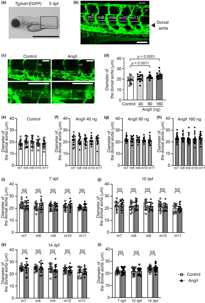
Angiotensin II (AngII) administration increased the dorsal aorta diameter in zebrafish embryos. (a) A lateral bright field image of Tg(kdrl:EGFP) control embryos at 5 days post‐fertilization (dpf). Scale bar: 1 mm. (b) A confocal microscopy image of the EGFP‐positive blood vessels of the boxed region in (a); m7 to m11 indicates myotomes 7–11, respectively. Scale bar: 100 μm. (c) Representative lateral dorsal aorta images of control or AngII (160 ng)‐injected Tg(kdrl:EGFP) embryos at 5 dpf. The lower panels are higher‐magnification images of the upper panels. Scale bars: 50 μm. (d) Quantification of the average diameter of the dorsal aorta. (e–h) Dorsal aorta diameter at myotomes 7–11 in control or AngII (40 ng, 80 ng, or 160 ng)‐injected Tg(kdrl:EGFP) embryos at 5 dpf. n = 15 (control), 15 (40 ng), 13 (80 ng), and 18 (160 ng). (i–k) Dorsal aorta diameter at myotomes 7–11 in control or AngII (160 ng)‐injected Tg(kdrl:EGFP) embryos at 7 dpf (i), 10 dpf (j), and 14 dpf (k). (l) Quantification of the average diameter of the five serial segments of the dorsal aorta. n = 14 (control, white), and n = 14 (AngII 160 ng, gray). NS indicates not significant. Dotted lines in (b) and (c) indicate the diameter of the dorsal aortae. DA, dorsal aorta; DLAV, dorsal longitudinal anastomotic vessel; ISV, intersegmental vessel; PCV, posterior cardinal vein.

Acknowledgments
This image is the copyrighted work of the attributed author or publisher, and ZFIN has permission only to display this image to its users. Additional permissions should be obtained from the applicable author or publisher of the image. Full text @ Physiol. Rep.