FIGURE SUMMARY
Title

Genetic evidence for differential functions of figla and nobox in zebrafish ovarian differentiation and folliculogenesis

Authors
Wu, K., Zhai, Y., Qin, M., Zhao, C., Ai, N., He, J., Ge, W.
Source
Full text @ Commun Biol

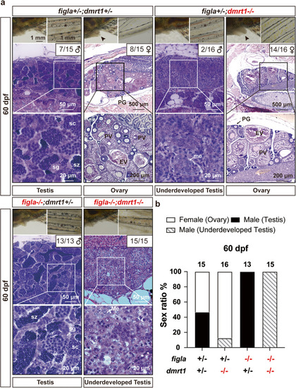
Phenotype analysis of different genotypes of figla and dmrt1 mutations at 60 dpf.

a Gonads and secondary sex characteristics of four different genotypes: normal ovary and testis in the control (figla+/−;dmrt1+/−; n = 15 independent fish, 7 males, 8 females); normal ovary and underdeveloped testis in dmrt1 single mutant (figla+/−;dmrt1−/−; n = 16 independent fish, 2 males, 14 females); all-male testis in figla single mutant (figla−/−;dmrt+/−; n = 13 independent fish, 13 males) and underdeveloped testis in figla and dmrt1 double mutant (figla−/−;dmrt1−/−; n = 15 independent fish). Asterisk, breeding tubercles on pectoral fins; arrowhead, genital papilla. b Sex ratio in four different genotypes at 60 dpf. The sample sizes for independent fish are shown at the top of the columns. PG primary growth, PV previtellogenic, EV early vitellogenic, me meiotic cells, sc spermatocytes, sg spermatogonia, sz spermatozoa.

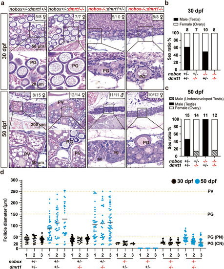
Rescue of the all-male phenotype of nobox mutant by simultaneous mutation of dmrt1.

a Gonadal histology of nobox mutant and double mutant with dmrt1 mutation in early gonadal differentiation (30 and 50 dpf). b Sex ratio in four different genotypes of nobox and dmrt1 mutations at 30 dpf (nobox+/−;dmrt1+/−: n = 8 independent fish; nobox+/−;dmrt1−/−: n = 7 independent fish; nobox−/−;dmrt1+/−: n = 10 independent fish; nobox−/−;dmrt1−/−: n = 8 independent fish). c Sex ratio in four different genotypes of nobox and dmrt1 mutations at 50 dpf (nobox+/−;dmrt1+/−: n = 15 independent fish; nobox+/−;dmrt1−/−: n = 14 independent fish; nobox−/−;dmrt1+/−: n = 11 independent fish; nobox−/−;dmrt1−/−: n = 12 independent fish). d Follicle composition of different genotypes at 30 and 50 dpf (n = 3 independent fish). The horizontal black lines represent the mean. PN perinucleolar oocytes, CN chromatin nucleolar oocytes.

Long-term degeneration of the double mutant ovaries.

a The ovaries in the double mutant (nobox−/−;dmrt1−/−) showed different degrees of degeneration at 120 dpf with loss of oocytes and decreased ovarian size (n = 5 independent fish). In addition, empty cavities or vacuoles left by degenerated oocytes were often observed (asterisks). The degenerated ovaries were gradually replaced by testicular tissues with meiotic cells (me); however, spermatogenesis could not proceed further due to the lack of dmrt1. The ovarian degeneration process was categorized into four stages based on gonadal size and morphological features. In stage I, the ovary was significantly smaller than the control (approximately half the size), containing both PG and PV follicles. Stage II was characterized by a dramatically reduced ovarian size, while still housing PG and PV follicles. In stage III, the ovary contained PG follicles only. In stage IV, the ovary was devoid of all follicles, featuring empty cavities or vacuoles left by degenerated oocytes. Additionally, testicular tissues began to emerge with meiotic germ cells. b Ovarian sizes of the control and double mutant ovaries undergoing degeneration. The area size of the largest section of each ovary was measured with ImageJ and the data are expressed as the ratios relative to the control. c Number of vacuoles resulting from oocyte loss. The vacuoles were counted on the largest section and classified based on their sizes compared to those of PG and PV follicles. d Composition of gonadal tissues in the control and double mutant ovaries. The areas of different gonadal tissues were measured using ImageJ.

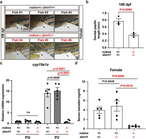
Evidence for estrogen deficiency in double mutant females.

a Genital papilla in the control (nobox+/−;dmrt1+/−; n = 3 independent fish) and double mutant (nobox−/−;dmrt1−/−; n = 3 independent fish) females (arrow). The dotted line showed the length of the genital papilla. b Length of genital papilla in the controls and double mutant females (n = 3 independent fish). Data shown are mean ± SEM (P = 0.0088 by unpaired Student’s two-tailed t test). c Expression of cyp19a1a in PG and PV follicles (n = 5 independent samples). Total RNA was extracted from the isolated PG and PV follicles and reverse transcribed into cDNA for real-time PCR analysis. Each data point represents PG or PV follicles isolated and pooled from two fish for each genotype. Data shown are mean ± SEM, P values revealed by one-way ANOVA and Tukey’s test. d Serum E2 levels in the control (nobox+/−;dmrt1+/−; n = 5 independent fish), dmrt1 single mutant (nobox+/−;dmrt1−/−; n =5 independent fish) and double mutant (nobox−/−; dmrt1−/−; n = 6 independent fish) females at 100 dpf. Data shown are mean ± SEM (nobox+/−;dmrt1+/− v.s nobox+/−;dmrt1−/−: P = 0.6649; nobox+/−;dmrt1+/− v.s nobox−/−;dmrt1−/−: P = 0.0095; nobox+/−;dmrt1−/− v.s nobox−/−;dmrt1−/−: P = 0.0018; P values revealed by one-way ANOVA and Tukey’s test).

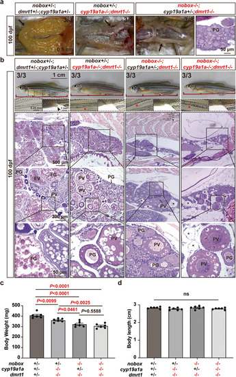
Ovarian growth and follicle development in the triple mutant of cyp19a1a, nobox and dmrt1.

a Gross morphology of the ovaries (arrow) in females of the control (nobox+/−;cyp19a1a+/−;dmrt1+/−, n = 3 independent fish), cyp19a1a and dmrt1 double mutant (nobox+/−;cyp19a1a−/−;dmrt1−/−, n = 3 independent fish), and nobox and dmrt1 double mutant (nobox−/−;cyp19a1a+/−;dmrt1−/−, n = 3 independent fish). b Ovarian histology and secondary sex characteristics in different genotypes: control (nobox+/−;cyp19a1a+/−;dmrt1+/−, n = 3 independent fish), cyp19a1a and dmrt1 double mutant (nobox+/−;cyp19a1a−/−;dmrt1−/−, n = 3), nobox and dmrt1 double mutant (nobox−/−;cyp19a1a+/−; dmrt1−/−, n = 3 independent fish), and nobox, cyp19a1a and dmrt1 triple mutant (nobox−/−;cyp19a1a−/−; dmrt1−/−, n = 3 independent fish). Red asterisk, breeding tubercles; arrowhead, genital papilla; black asterisk, vacuoles left by the lost oocytes. c, d Body weight and body length of the triple mutant of cyp19a1a, nobox and dmrt1 (n = 6 independent fish). Data shown are mean ± SEM, P values revealed by one-way ANOVA and Tukey’s test.

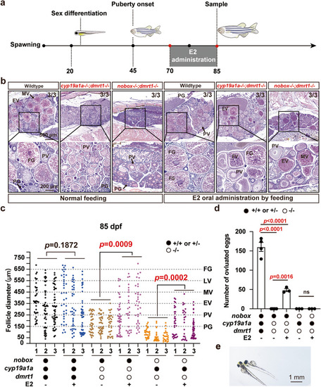
Rescue of vitellogenic growth in the double mutants (
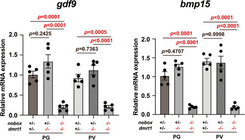
nobox−/−;dmrt1−/− and cyp19a1a−/−;dmrt1−/−) by E2 treatment.

a Schematic illustration of E2 treatment. b Ovarian histology of different genotypes in the control group with normal feeding and the E2 treatment group fed with an E2-containing diet (n = 3 independent fish). Vitellogenic growth characterized with yolk granule accumulation resumed in both double mutants (nobox−/−;dmrt1−/− and cyp19a1a−/−;dmrt1−/−), which were blocked at the PV stage with the formation of cortical alveoli but not yolk granules. c Follicle composition of different genotypes in different treatment groups (n = 3 independent fish). The data points shown are diameters of individual follicles and the statistical significance of the means was demonstrated by unpaired Student’s two-tailed t test. d Fecundity of different genotypes and treatments. Each data point represents the number of eggs spawned by each female mated with one wild-type male (Control group: n = 4 independent experiments; Other groups: n = 3 independent experiments). The sexes of examined females were further confirmed by histology after mating. Data shown are mean ± SEM, P values revealed by one-way ANOVA and Tukey’s test. e The offspring from E2-treated females of cyp19a1a−/−;dmrt1−/− double mutant.

Expression of gdf9 and bmp15 genes in PG and PV follicles of nobox−/−;dmrt1−/− double mutant at 100 dpf.

Total RNA was extracted from the isolated PG and PV follicles and reverse transcribed into cDNA for real-time PCR analysis. Each data point represents PG or PV follicles isolated and pooled from two fish for each genotype, totaling 10 fish (n = 5 independent samples). The mRNA levels of each target gene were normalized to that of the housekeeping gene ef1a, and expressed as a fold change relative to the control. Data shown are mean ± SEM, P values revealed by one-way ANOVA and Tukey’s test.

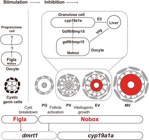
A working model on differential roles of Figla and Nobox in zebrafish folliculogenesis.

Figla plays a critical role in controlling follicle formation in the event of cyst breakdown or follicle assembly, while Nobox is more involved in regulating follicle development after its formation, including such events as follicle activation (PG-PV transition) and vitellogenic growth (PV-EV transition). Nobox controls vitellogenic growth by regulating aromatase (cyp19a1a) expression in the follicle cells, which may be mediated by oocyte-secreted signaling molecules such as Gdf9 and Bmp15.

Acknowledgments
This image is the copyrighted work of the attributed author or publisher, and ZFIN has permission only to display this image to its users. Additional permissions should be obtained from the applicable author or publisher of the image. Full text @ Commun Biol