FIGURE SUMMARY
Title

A novel role for the chloride intracellular channel protein Clic5 in ciliary function

Authors
Ott, E., Hoff, S., Indorf, L., Ditengou, F.A., Müller, J., Renschler, G., Lienkamp, S.S., Kramer-Zucker, A., Bergmann, C., Epting, D.
Source
Full text @ Sci. Rep.

Expression analyses of clic5 in zebrafish. (A) Semi-quantitative reverse-transcription polymerase chain reaction (RT-PCR) on cDNA of different adult zebrafish organs revealed expression of clic5a (223 bp) in the spleen and of clic5b (408 bp) in the gut, kidney and liver. H2O served as negative control and ef1α as loading control; Primer dimer (PD). (B) Whole-mount in situ hybridization (WISH) analysis reveals expression of clic5 in the pronephric tubules (black arrow) at 1 day post-fertilization (dpf) which persisted throughout the first days of zebrafish development. At 2dpf, clic5 was additionally expressed in the eye lenses (black arrowheads), liver primordium (white arrowhead), pectoral fin buds and glomerulus (white arrows). At 5dpf clic5 expression was detectable in the liver (white arrowhead) and gut (black arrowheads) (framed box shows the expression of clic5 in the liver and gut from the dorsal view). WISH analysis with specific probes for clic5a and clic5b reveals expression of clic5a in the eye lenses and glomerulus at 2dpf and expression of clic5b in the pronephric tubules at 1 and 2dpf with additional expression in the pectoral fin buds and liver primordium at 2dpf.

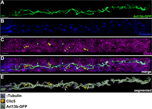
Subcellular localization analyses of Clic5 in the pronephric tubule of zebrafish. (A–D) Triple whole-mount immunostaining on 1dpf old Tg(actb2:Mmu.Arl13b-GFP) zebrafish embryos using anti-GFP (labeling Arl13b-GFP, i.e. the ciliary axoneme) (A), anti-γTubulin (labeling the ciliary basal body) (B) and anti-Clic5 (monoclonal antibody produced in mouse) (C) antibodies reveals ciliary localization of Clic5 in the pronephric tubule. (D) Image is (A–C) merged. (E) 3D reconstruction and segmentation of Arl13b-GFP (green), γTubulin (blue) and Clic5 (gold) using Imaris software. Clic5 voxels co-localize with those of ciliary basal body (white arrows) and ciliary axoneme voxels (yellow arrows), respectively. Scale bar, 5 µm.

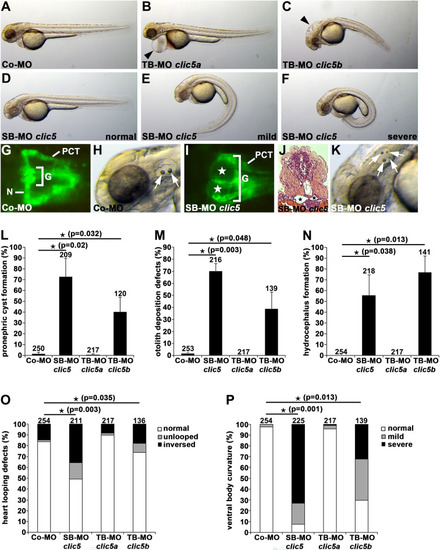
Clic5 knockdown analyses of cilia-related phenotypes in zebrafish. (A–F) Bright-field images of zebrafish embryos at 2dpf injected with Co-MO (6 ng) (A), translation-blocking morpholino (TB-MO) clic5a (6 ng) (B), TB-MO clic5b (4 ng) (C) and splicing-blocking morpholino (SB-MO) clic5 (4 ng) (D–F). In comparison to Co-MO injected embryos, injection of TB-MO clic5a leads to pericardial edema (black arrowhead) and injection of TB-MO clic5b or SB-MO clic5 leads to hydrocephalus formation (black arrowhead) and different degrees of ventral body curvature. (G–K) Knockdown of Clic5 leads to pronephric cyst formation (white and black stars in (I,J), respectively) and otolith deposition defects (white arrows) at 2dpf as shown in a dorsal view with anterior to the left of a SB-MO clic5 injected Tg(wt1b:EGFP) embryo (glomerulus (G), neck (N), proximal convoluted tubule (PCT)) (I), and an embryo shown in a bright-field image (K), respectively, in comparison to Co-MO injected embryos (G,H); histological transverse section reveals pronephric cyst formation in a SB-MO clic5 injected embryo (J). (L–P) Quantification of pronephric cyst formation (L), otolith deposition defects (M), hydrocephalus formation (N), altered heart looping (O) and ventral body curvature (P) in 2dpf zebrafish embryos injected with Co-MO (6 ng), SB-MO clic5 (4 ng), TB-MO clic5a (6 ng) or TB-MO clic5b (4 ng).

Analyses of Clic5 in ciliogenesis, and in ciliary and glomerular function in zebrafish. (A) Anti-acetylated Tubulin immunostaining reveals reduced cilia formation in the pronephric tubules of embryos injected with SB-MO clic5 (4 ng) or TB-MO clic5b (4 ng) in comparison to Co-MO (4 ng) injected embryos at 1dpf. Representative confocal images depict the middle part of the pronephric tubule for each condition with anterior to the left. Scale bar: 10 µm. (B) Quantitative RT-PCR analyses reveals unaltered expression of Hedgehog signalling components gli1 and ptc1 while Wnt signalling components axin2, wnt8a and lef1 were upregulated upon SB-MO clic5 (4 ng) mediated knockdown compared to the control at 1dpf. (C,D) Injection of TB-MO clic5a (6 ng) leads to detectable FITC-dextran 500 kDa in the pronephric tubules at 4dpf of zebrafish development in comparison to Co-MO (6 ng) injected embryos shown by respective confocal images of Tg(cdh17:mcherry) embryos. Scale bar: 10 µm. (E) Quantification reveals statistic significant FITC-dextran 500 kDa positive embryos for the knockdown of Clic5 and Clic5a compared to the knockdown of Clic5b or the control.

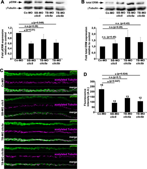
Analyses of pERM and total ERM levels after knockdown of Clic5, Clic5a or Clic5b. (A) Immunoblot analyses and respective quantification reveals significant decreased pERM levels in whole protein lysates from 1dpf old embryos that were injected with SB-MO clic5 (2 ng) or TB-MO clic5b (2 ng) compared to TB-MO clic5a (3 ng) or Co-MO (3 ng) injected embryos. (B) Immunoblot analyses and respective quantification of total ERM levels reveals no significant difference in whole protein lysates from 1dpf old embryos that were injected with SB-MO clic5 (2 ng), TB-MO clic5a (3 ng) or TB-MO clic5b (2 ng) compared with Co-MO (3 ng) injected embryos. (C,D) Double whole-mount immunostaining and respective quantification of 1dpf old zebrafish embryos using anti-pERM (green) and anti-acetylated Tubulin (magenta) antibodies reveals significant reduced pERM levels in the pronephric tubules of embryos injected with SB-MO clic5 (2 ng) or TB-MO clic5b (2 ng) compared to TB-MO clic5a (3 ng) and Co-MO (3 ng) injected embryos; Arbitrary Unit (A.U.). Scale bar: 10 µm.

Acknowledgments
This image is the copyrighted work of the attributed author or publisher, and ZFIN has permission only to display this image to its users. Additional permissions should be obtained from the applicable author or publisher of the image. Full text @ Sci. Rep.