IMAGE

Figure 2

ID
ZDB-IMAGE-231019-4
Source
Figures for Ott et al., 2023
Image
Figure Caption

Figure 2

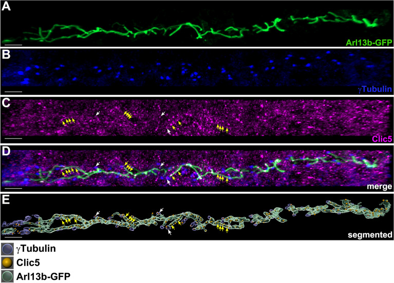
Subcellular localization analyses of Clic5 in the pronephric tubule of zebrafish. (A–D) Triple whole-mount immunostaining on 1dpf old Tg(actb2:Mmu.Arl13b-GFP) zebrafish embryos using anti-GFP (labeling Arl13b-GFP, i.e. the ciliary axoneme) (A), anti-γTubulin (labeling the ciliary basal body) (B) and anti-Clic5 (monoclonal antibody produced in mouse) (C) antibodies reveals ciliary localization of Clic5 in the pronephric tubule. (D) Image is (A–C) merged. (E) 3D reconstruction and segmentation of Arl13b-GFP (green), γTubulin (blue) and Clic5 (gold) using Imaris software. Clic5 voxels co-localize with those of ciliary basal body (white arrows) and ciliary axoneme voxels (yellow arrows), respectively. Scale bar, 5 µm.

Acknowledgments
This image is the copyrighted work of the attributed author or publisher, and ZFIN has permission only to display this image to its users. Additional permissions should be obtained from the applicable author or publisher of the image. Full text @ Sci. Rep.