IMAGE

Figure 3

ID
ZDB-IMAGE-231019-5
Source
Figures for Ott et al., 2023
Image
Figure Caption

Figure 3

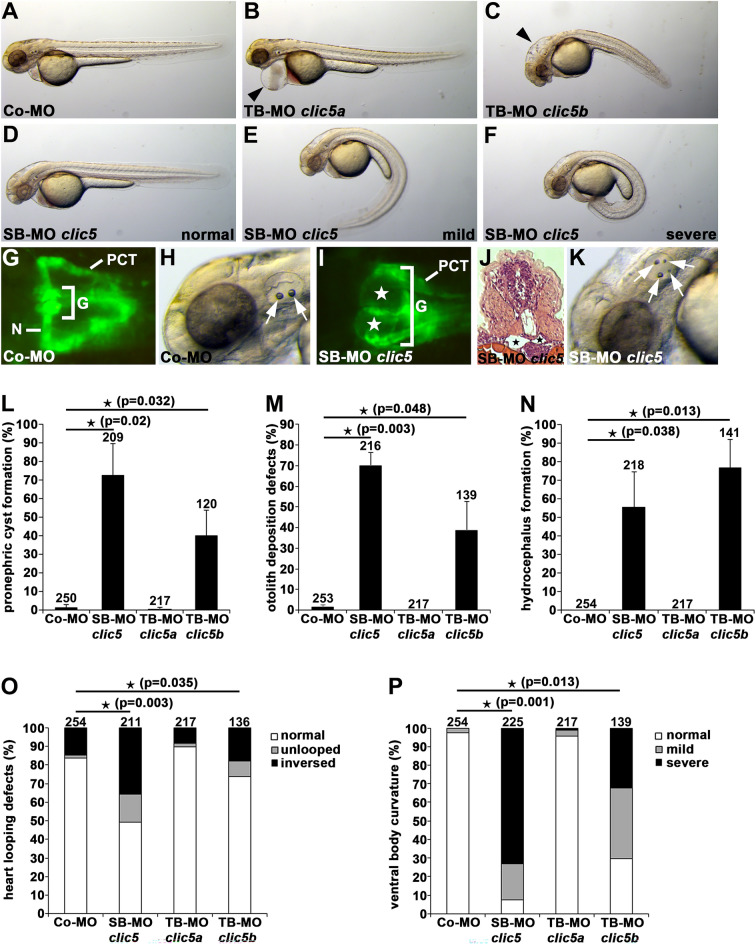
Clic5 knockdown analyses of cilia-related phenotypes in zebrafish. (A–F) Bright-field images of zebrafish embryos at 2dpf injected with Co-MO (6 ng) (A), translation-blocking morpholino (TB-MO) clic5a (6 ng) (B), TB-MO clic5b (4 ng) (C) and splicing-blocking morpholino (SB-MO) clic5 (4 ng) (D–F). In comparison to Co-MO injected embryos, injection of TB-MO clic5a leads to pericardial edema (black arrowhead) and injection of TB-MO clic5b or SB-MO clic5 leads to hydrocephalus formation (black arrowhead) and different degrees of ventral body curvature. (G–K) Knockdown of Clic5 leads to pronephric cyst formation (white and black stars in (I,J), respectively) and otolith deposition defects (white arrows) at 2dpf as shown in a dorsal view with anterior to the left of a SB-MO clic5 injected Tg(wt1b:EGFP) embryo (glomerulus (G), neck (N), proximal convoluted tubule (PCT)) (I), and an embryo shown in a bright-field image (K), respectively, in comparison to Co-MO injected embryos (G,H); histological transverse section reveals pronephric cyst formation in a SB-MO clic5 injected embryo (J). (L–P) Quantification of pronephric cyst formation (L), otolith deposition defects (M), hydrocephalus formation (N), altered heart looping (O) and ventral body curvature (P) in 2dpf zebrafish embryos injected with Co-MO (6 ng), SB-MO clic5 (4 ng), TB-MO clic5a (6 ng) or TB-MO clic5b (4 ng).

Acknowledgments
This image is the copyrighted work of the attributed author or publisher, and ZFIN has permission only to display this image to its users. Additional permissions should be obtained from the applicable author or publisher of the image. Full text @ Sci. Rep.