FIGURE SUMMARY
Title

kcnj13 regulates pigment cell shapes in zebrafish and diverged by cis-regulatory evolution between Danio species

Authors
Podobnik, M., Singh, A.P., Fu, Z., Dooley, C.M., Frohnhöfer, H.G., Firlej, M., Stednitz, S.J., Elhabashy, H., Weyand, S., Weir, J.R., Lu, J., Nüsslein-Volhard, C., Irion, U.
Source
Full text @ Development

Pigment patterns in wild-type and kcnj13 mutant D. rerio and D. aesculapii. (A-H) Pigment patterns in D. rerio wild type (A), D. aesculapii wild type (B), D. rerio kcnj13t24ui (C), D. aesculapii kcnj13t11mp (D), D. rerio kcnj13txg6 (E) and D. rerio kcnj13td15 (F). kcnj13t24ui and kcnj13td15 were crossed to produce trans-heterozygous kcnj13tui24/td15 F1 fish (not shown), which were then incrossed to generate F2 fish with the genotypes kcnj13t24ui (G; n=8) and kcnj13td15 (H; n=12). (I-M) Melanophore clearance in kcnj13tdxg6 is similar to wild type during the development of the first light stripe between 25 and 45 dpf (n=2). (N-O′) D. rerio wild-type (N,N′) and kcnj13tdxg6 (O,O′) patterns at juvenile stage (J), 11 mm standard length (SL; for staging see Materials and Methods) (n>10). In the mutants, iridophores fail to reiterate the consecutive light stripes, which ultimately leads to fewer and broader stripes with occasional interruptions. Light and dark grey bars represent light and dark stripe areas, respectively. Stripes are denoted as in Parichy et al. (2009).

Melanophores require kcnj13 autonomously during stripe formation. (A-C) Testing cell-autonomy of kcnj13 by blastula transplantations reveals a genetic requirement in melanophores (A; kcnj13td15;ednrbatlf802;csf1ratm236b into mitfaw2; n=1), but not in xanthophores (B; kcnj13td15;kitab134;ednrbatlf802 into csf1ratm236b; n=2) or iridophores (C; kcnj13td15;mitfaw2;csf1ratm236b into ednrbatlf802; n=1). (D-D″) Transplantation experiments (kcnj13tui24 into slc45a2b4) provide further evidence of a cell-autonomous function of kcnj13 in melanophores during stripe formation. Transplanted mutant melanophores (pigmented) are associated with stripe perturbations in slc45a2 hosts (n=3). Strong pattern deformations are never observed in chimeras without pigmented trunk melanophores (n=41). Control transplantations of wild-type melanophores into slc45a2b4 hosts do not cause such pattern deformations (Dooley et al., 2013a,b).

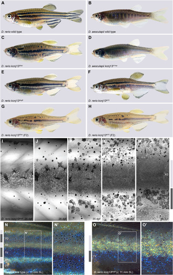
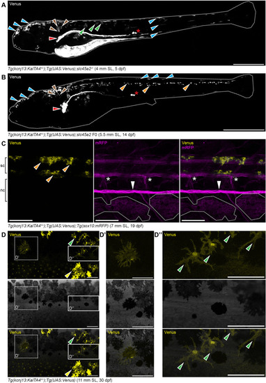
Endogenous kcnj13 expression during D. rerio development. (A) Heterozygous KalTA4::Venus reporter larva (n>50) showing signals in melanophores in the head and tail regions (cyan arrowheads), xanthophores (green arrowheads), hindbrain (brown arrowheads), along the entire pronephros (red arrowhead), including corpuscles of Stannius (red asterisk), and the yolk. 4 mm standard length (SL), 5 dpf, sagittal view, images of four positions along the A-P axis combined into one composite. (B) Similar expression patterns can be observed in larva 1 week older (n>25), with additional signals in the spinal cord (orange arrowheads). These signals persist throughout further development. 5.5 mm SL, 14 dpf, sagittal view, images of five combined into one composite. (C) Venus expression does not overlap with locations of the pigment cell stem cells at the DRGs (marked by white asterisks) in reporter larva (n>10). Iridophore patches in the skin indicated with white dotted outlines, lateral line nerve marked with a white arrowhead; presumptive neurons in the spinal cord marked with orange arrowheads. nc, notochord; sc, spinal cord. 7 mm SL, 19 dpf. (D-D″) During and after the consolidation of the stripes in wild types (n>10, see Fig. 1I-O), Venus expression can be detected in only a minority of melanophores (D′) and xanthophores (D″) in the skin at any given time point. Green arrowheads indicate stellate and Venus-positive xanthophores in the dark stripe, yellow arrowheads indicate compact, pigmented and Venus-positive xanthophores in the light stripe. 11 mm SL, 30 dpf. Scale bars: 500 µm (A); 1 mm (B); 100 µm (C-D″).

Pigment cell organization and shapes in D. rerio wild types and kcnj13 mutants, and D. aesculapii wild types. (A) In adult wild-type D. rerio (n>3), melanophores in the stripe are densely packed (note variegation of the transgene in a few cells indicated with light-grey arrowheads) and cells at the boundary form long protrusions towards the light stripe (cyan arrowheads). (B) In kcnj13tdxg6 mutants (n=2), cells are less tightly packed in the dark stripe and short protrusions form without clear polarity (cyan arrowheads). (C) Wild-type xanthophores acquire stellate shapes in the dark stripes (green arrowheads) and compact shapes in the light stripes (yellow arrowheads) (n>3, Mahalwar et al., 2014). (D) Transplanted mRFP-positive wild-type xanthophores acquire inappropriate compact shapes (yellow arrowheads) in dark stripes in kcnj13tdxg6 mutants [donor: Tg(sox10:mRFP), host: kcnj13tdxg6; n=2]. In control transplantations labelled wild-type xanthophores acquire loose shapes in the dark stripes in wild types (Mahalwar et al., 2014). (E) Wild-type iridophores acquire loose shapes (white arrowheads) in the dark stripes and dense shapes (magenta arrowheads) in the light stripes (n>3, Singh et al., 2014). (F) Iridophores acquire ectopic compact shapes (magenta arrowheads) in the dark stripes in kcnj13tdxg6 mutants (n>2), visualized by tracing labelled clones. Light and dark grey bars represent light and dark stripes in D. rerio, respectively. (G) Wild-type D. rerio form long melanophore protrusions towards the light stripe regions (cyan arrowheads, see A; n>3). (H) Melanophore protrusions are not polarized in D. rerio kcnj13 mutants (cyan arrowheads, see B; n>3) and pigmented xanthophores are visible in the dark stripe region (yellow arrowheads). (I) D. aesculapii wild types lack polarized melanophores (cyan arrowheads), melanophores and xanthophores mix occasionally, and the boundary between bars and light regions is of very low contrast (n>3). lr, light region; mb, melanophore bar region. Scale bars: 100 µm (A,B); 250 µm (C-F); 500 μm (G-I).

Molecular basis of kcnj13 evolution between D. rerio and D. aesculapii. (A-D) D. rerio wild type (A) and D. rerio kcnj13t24ui (B), in which either the D. rerio allele of kcnj13 [C; Tg(mitfa:kcnj13D.rerio);kcnj13tui24; n>50] or the D. aesculapii kcnj13 allele [D; Tg(mitfa:kcnj13D.aesculapii);kcnj13tui24; n>150] was expressed under the control of the mitfa promoter from D. rerio. In both cases, stripes were restored in the trunk of the fish. R224K was found to be polymorphic in D. aesculapii (Podobnik et al., 2020). (E) SWISS-MODEL derived homology model of the Kcnj13 tetramer (Q23L and D180G diverged between species in magenta). (F) Allele-specific transcriptome analysis, based on the D. rerio reference genome, shows higher kcnj13 expression of the D. rerio allele in the skin of interspecific hybrids (n=12; P-adjust<0.001). A similar expression bias is observed in the trunk of the hybrids (Fig. S3A); analysis of the same RNA-seq data using the D. aesculapii genome as reference yielded very similar results (Fig. S3B,C).

Acknowledgments
This image is the copyrighted work of the attributed author or publisher, and ZFIN has permission only to display this image to its users. Additional permissions should be obtained from the applicable author or publisher of the image. Full text @ Development