IMAGE

Fig. 4.

ID
ZDB-IMAGE-230916-286
Source
Figures for Podobnik et al., 2023
Image
Figure Caption

Fig. 4.

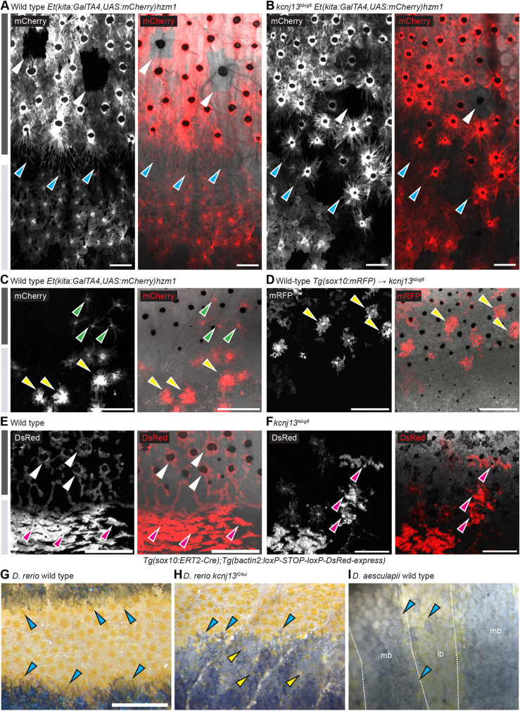
Pigment cell organization and shapes in D. rerio wild types and kcnj13 mutants, and D. aesculapii wild types. (A) In adult wild-type D. rerio (n>3), melanophores in the stripe are densely packed (note variegation of the transgene in a few cells indicated with light-grey arrowheads) and cells at the boundary form long protrusions towards the light stripe (cyan arrowheads). (B) In kcnj13tdxg6 mutants (n=2), cells are less tightly packed in the dark stripe and short protrusions form without clear polarity (cyan arrowheads). (C) Wild-type xanthophores acquire stellate shapes in the dark stripes (green arrowheads) and compact shapes in the light stripes (yellow arrowheads) (n>3, Mahalwar et al., 2014). (D) Transplanted mRFP-positive wild-type xanthophores acquire inappropriate compact shapes (yellow arrowheads) in dark stripes in kcnj13tdxg6 mutants [donor: Tg(sox10:mRFP), host: kcnj13tdxg6; n=2]. In control transplantations labelled wild-type xanthophores acquire loose shapes in the dark stripes in wild types (Mahalwar et al., 2014). (E) Wild-type iridophores acquire loose shapes (white arrowheads) in the dark stripes and dense shapes (magenta arrowheads) in the light stripes (n>3, Singh et al., 2014). (F) Iridophores acquire ectopic compact shapes (magenta arrowheads) in the dark stripes in kcnj13tdxg6 mutants (n>2), visualized by tracing labelled clones. Light and dark grey bars represent light and dark stripes in D. rerio, respectively. (G) Wild-type D. rerio form long melanophore protrusions towards the light stripe regions (cyan arrowheads, see A; n>3). (H) Melanophore protrusions are not polarized in D. rerio kcnj13 mutants (cyan arrowheads, see B; n>3) and pigmented xanthophores are visible in the dark stripe region (yellow arrowheads). (I) D. aesculapii wild types lack polarized melanophores (cyan arrowheads), melanophores and xanthophores mix occasionally, and the boundary between bars and light regions is of very low contrast (n>3). lr, light region; mb, melanophore bar region. Scale bars: 100 µm (A,B); 250 µm (C-F); 500 μm (G-I).

Acknowledgments
This image is the copyrighted work of the attributed author or publisher, and ZFIN has permission only to display this image to its users. Additional permissions should be obtained from the applicable author or publisher of the image. Full text @ Development