
- Title
-
A novel insight on SARS-CoV-2 S-derived fragments in the control of the host immunity
- Authors
- Bastos, T.S.B., de Paula, A.G.P., Dos Santos Luz, R.B., Garnique, A.M.B., Belo, M.A.A., Eto, S.F., Fernandes, D.C., Ferraris, F.K., de Pontes, L.G., França, T.T., Barcellos, L.J.G., Veras, F.P., Bermejo, P., Guidelli, G., Maneira, C., da Silveira Bezerra de Mello, F., Teixeira, G., Pereira, G.A.G., Fernandes, B.H.V., Sanches, P.R.S., Braz, H.L.B., Jorge, R.J.B., Malafaia, G., Cilli, E.M., Olivier, D.D.S., do Amaral, M.S., Medeiros, R.J., Condino-Neto, A., Carvalho, L.R., Machado-Santelli, G.M., Charlie-Silva, I., Galindo-Villegas, J., Braga, T.T.
- Source
- Full text @ Sci. Rep.
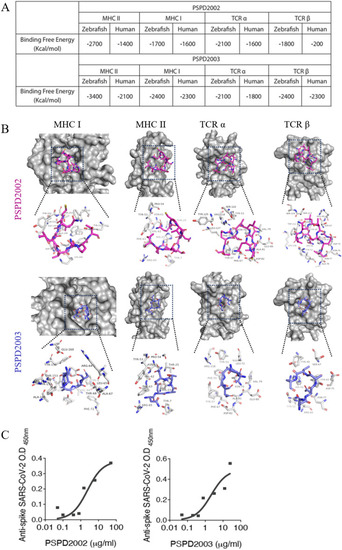
Peptides PSPD2002 and PSPD2003 potentially interact with receptors of the human and zebrafish immune systems. (A) Calculated interaction free energy (Kcal/mol) for PSPD2002 and PSPD2003 interactions for MHC II, MHC class I, TCRα and TCRβ in Homo sapiens and Danio rerio homologous receptors. (B) Docking analysis highlighting the interaction sites between PSPD2002 (purple) and PSPD2003 (blue), and the MHC II, MHC I, TCRα and TCRβ ligands of Danio rerio. Serial dilutions of the peptides being recognized by anti-Spike SARS-CoV-2 antibody for evaluation of the specificity of PSPD2002, in (C), and PSPD2003, in (D). |
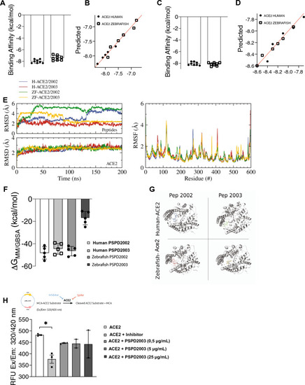
Affinity and binding stability between S-derived peptides and the ACE2 receptor. (A) and (C) Molecular dynamics analysis of interaction strength between PSPD2002 (A) or PSPD2003 (C) and ACE2 receptor of human and zebrafish, and predictive values for PSPD2002 (B) or PSPD2003 (D). (E) Analysis of the convergence and stability of the interaction of proteases over 200 ns for peptides (top) and ACE2 (bottom), from RMSD (root-mean-square deviation) analysis (left). Fluctuation analysis of the different residues of the proteases using RMSF (root-mean-square fluctuation) docking (right). (F) Analysis of the binding free energy during 100 ns of stimulation, between both peptides (PSPD2002 and PSPD2003) and the human and zebrafish Ace2 receptors. (G) Graphical representation showing the interaction regions between the PSPD2002 and PSPD2003 fragments with the proteases of the quaternary structure of the human and zebrafish Ace2 receptors. (H) Expression of ACE2 in Saccharomyces cerevisiae strain. The analysis of the catalytic activity of the receptor against the synthetic inhibitor (control) and different concentrations of the peptides was quantified from the Relative Fluorescence Unit (RFU) read in excitation range of 320 nm and emission range of 420 nm (Ex/Em: 320/420). Å: angstrom, a unit of length equivalent to 0.1 nm. MM/GBSA: Molecular mechanics generalized Born surface area. *p < 0.05. |
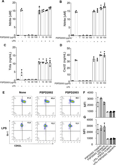
Effect of PSPD2002 and PSPD2003 peptides on mammalian cells. Quantification of nitric oxide (NO) production in AMJ2-C11 cells in the presence and absence of lipopolysaccharide (LPS), challenged with 1, 10, 30 and 100 ug/ml of PSPD2002 (A) and PSPD2003 (B). Quantification of Tnfa (C) and Cxcl2 (D), in AMJ2-C11 cells challenged with PSPD2003, in the presence and absence of LPS. E Neutrophils (GR1+ cells) activation status demonstrated by representative flow cytometry graphics of unsensitized (top row) or sensitized with LPS (bottom row) and challenged with PSPD2002 (middle column) or PSPD2003 (right column). The x-axis represents L-selectin (CD62L) marker. (F) and (G) Mean fluorescence intensity (MFI) quantification of cells unsensitized (F) or sensitized with LPS (G). All experiments were performed in triplicate in three independent days. All data are expressed as the mean ± standard deviation. p < 0.05 was considered to indicate a statistically significant difference. |
Effects of immunization of S-derived peptides on zebrafish larvae. (A) Kaplain-Meyer curve representing the survival rate (y-axis) over 10 days (x-axis) of larvae immunized with water, and with different concentration (1 or 10 µg/mL) of PSPD2002 and PSPD2003. (B) Table with the main histopathological changes observed in zebrafish larvae immunized with PSPD2002 and PSPD2003. Different organs from different systems were analyzed and classified according to the alterations/pathology in a score ranging from 0 to 10. (C) Representative images obtained in confocal microscope of transgenic Tg(mpeg:mCherry/tnfa:eGFP) larvae immunized by inoculation into the swim bladder with different concentrations of PSPD2002 and PSPD2003 at 1, 2 and 6 days post injection (dpi). Macrophages are marked by red fluorescence (mCherry) expression and tnfa by green fluorescent protein (GFP) expression. The region surrounded by the yellow dash represents the counting region. Lateral view in sagittal plane of the animal, with caudal-cranial orientation (left–right) and pelvic fin below, allowing visualization of the swim bladder (centered), intestinal tract (below), and notochord and muscle tissue above. Counting the total number of macrophages (mCherry+) (D) and double-labeled pro-inflammatory macrophages (mCherry + /tnfa +) (E) in larvae immunized with PSPD2002 at 1, 2 and 6dpi. Counting the total number of macrophages (mCherry +) (F) and dual labeled pro-inflammatory macrophages (mCherry + /tnfa +) (G) in larvae immunized with PSPD2003 at 1, 2 and 6dpi. Scale bar 100um in (C) From C to G, experiments were performed in three independent days and n vary from 4 to 12. All data are expressed as the mean ± standard deviation. *p < 0.05; **p < 0.01. |
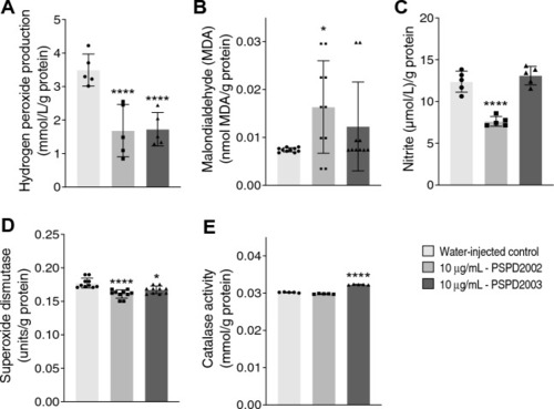
Changes in biomarkers of oxidative stress in zebrafish larvae immunized with 10 ug/ml of PSPD2002 or PSPD2003. The production levels of the oxidant compounds: hydrogen peroxide (A), malondialdehyde (B), nitrite (C), and of the antioxidants: superoxide dismutase (D) and catalase (E) were quantified. Experiments were performed in three independent days and n vary from 5 to 10. All data are expressed as the mean ± standard deviation. *p < 0.05; ****p < 0.0001 in the comparison with water-injected control. |