
Figure 3
- ID
- ZDB-FIG-230520-3
- Publication
- Bastos et al., 2023 - A novel insight on SARS-CoV-2 S-derived fragments in the control of the host immunity
- Other Figures
- All Figure Page
- Back to All Figure Page
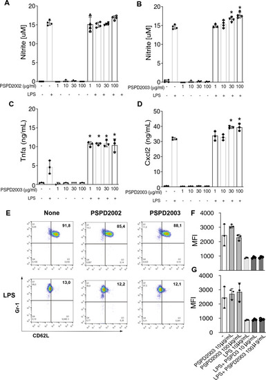
Effect of PSPD2002 and PSPD2003 peptides on mammalian cells. Quantification of nitric oxide (NO) production in AMJ2-C11 cells in the presence and absence of lipopolysaccharide (LPS), challenged with 1, 10, 30 and 100 ug/ml of PSPD2002 (A) and PSPD2003 (B). Quantification of Tnfa (C) and Cxcl2 (D), in AMJ2-C11 cells challenged with PSPD2003, in the presence and absence of LPS. E Neutrophils (GR1+ cells) activation status demonstrated by representative flow cytometry graphics of unsensitized (top row) or sensitized with LPS (bottom row) and challenged with PSPD2002 (middle column) or PSPD2003 (right column). The x-axis represents L-selectin (CD62L) marker. (F) and (G) Mean fluorescence intensity (MFI) quantification of cells unsensitized (F) or sensitized with LPS (G). All experiments were performed in triplicate in three independent days. All data are expressed as the mean ± standard deviation. p < 0.05 was considered to indicate a statistically significant difference. |