
- Title
-
Impaired fin regeneration and angiogenesis in aged zebrafish and turquoise killifish
- Authors
- Örling, J., Kosonen, K., Villman, J., Reichard, M., Paatero, I.
- Source
- Full text @ Biol. Open
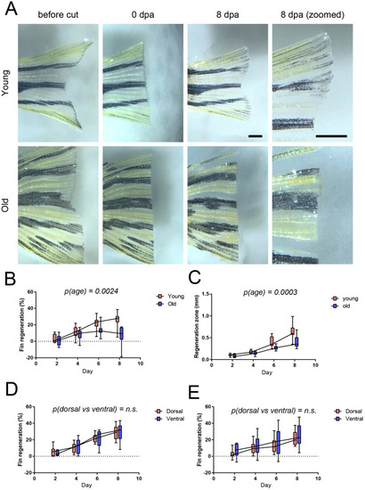
Aging reduces the regeneration of zebrafish caudal fin. Old (4 years) and young (4 months) male zebrafish were subjected to caudal fin regeneration assay. (A) Microscopy images of fins before, right after and 8 days after resection. Scale bar: 1 mm. (B) Quantification of the total regeneration of the caudal fin. Two-way ANOVA with age and time as factors was used for statistical analysis. All time points were used in the statistical analysis. (C) Quantitation of thickness of regenerating area. Two-way ANOVA with age and time as factors was used for statistical analysis. All time points were used in the statistical analysis. (D,E) Comparison of the regeneration in dorsal and ventral part of caudal fin. Data from young fish in panel D and in old fish in E. n=12 in both age groups. Two-way ANOVA, with age and anatomical location as factors, was used for statistical analysis. |
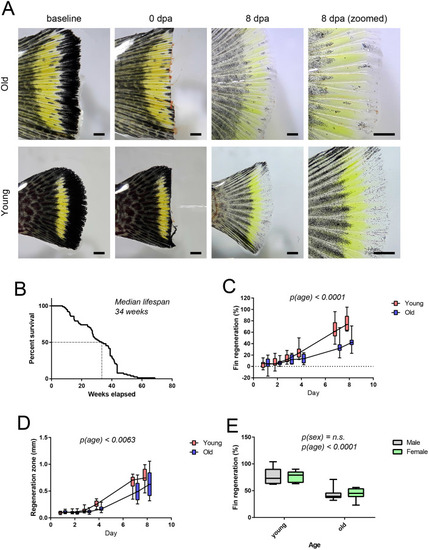
Aging reduces the regeneration of turquoise killifish caudal fin. Old and young turquoise killifish of both sexes were subjected to caudal fin regeneration assay. In total four groups were used (young male, old male, young female, old female). Young were 5 weeks of age and old 12 weeks when entering the experiment. (A) Microscopy images of turquoise killifish caudal fins before, immediately after and 8 days after resection. Scale bar: 1 mm. (B) Kaplan–Meier survival curve of turquoise killifish strain MSCZ222 housed in Turku, Finland. n=77. (C) Quantification of the total regeneration of the caudal fin. Two-way ANOVA with age and time as factors was used for statistical analysis, n=8 per group. All time points were used in the statistical analysis. (D) Thickness of regenerating zone. Two-way ANOVA with age and time as factors was used for statistical analysis. All timepoints were used in the statistical analysis. (E) Comparison of the total regeneration in male and female turquoise killifish caudal fin. In E, two-way ANOVA with sex and age as factors was used for statistical analysis. n=8 per group. |
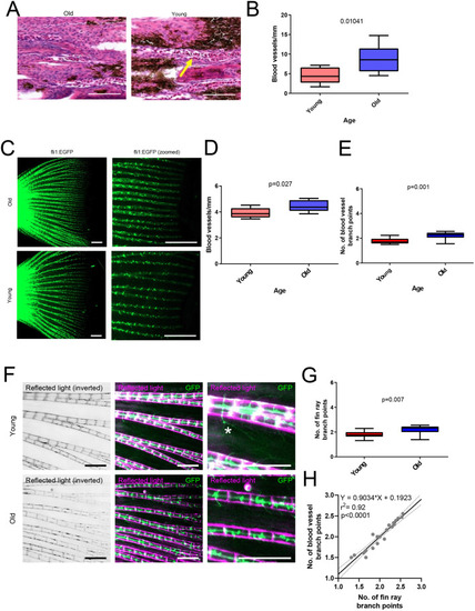
Aging increases vascular density in caudal fins. (A) Images of blood vessels in histological section of old and young turquoise killifish caudal fins stained with Hematoxylin and Eosin. An example of a blood vessel is indicated with a yellow arrow. Scale bar: 100 µm. (B) Quantification of blood vessel density in turquoise killifish sections. Statistical analysis with Mann–Whitney U-test, n=8 per group. (C) Fluorescence images of vasculature of transgenic zebrafish [roy, mitfa, Tg(fli1:EGFP)] caudal fins. Old fish were 36 months (n=8) and young fish 16 months (n=10). Scale bar: 1 mm. (D) Quantification of blood vessel density in transgenic zebrafish caudal fins. Statistical analysis with Mann–Whitney U-test. (E) Quantification of major blood vessel branch points in transgenic zebrafish caudal fins. (F) Fluorescence and reflected white light illumination images of transgenic zebrafish caudal fins. Scale bar: 0.5 mm. (G) Quantification of branching of fin rays. (H) Correlation analysis of fin ray and vascular branches. Statistical analysis using linear regression, both old and young samples combined, n=18. |
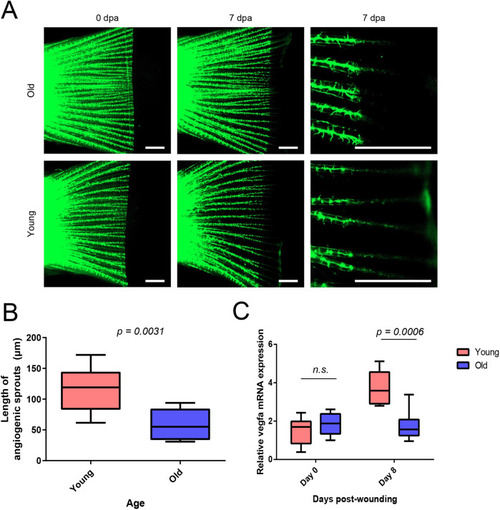
Angiogenesis is reduced in aged regenerating caudal fins. Zebrafish [genotype roy, mitfa, Tg(fli1:EGFP)] were subjected to caudal fin angiogenesis assay. Old fish were 36 months of age and young fish 16 months. (A) Fluorescence microscopy images of transgenic zebrafish caudal fins during fin regeneration experiment. Statistical analysis with Mann–Whitney U-test. (B) Quantification of angiogenesis in transgenic zebrafish caudal fins. Old fish (n=8), young fish (n=10). (C) qRT-PCR analysis of vegfa mRNA expression during fin regeneration assay in turquoise killifish. n=6 in each group. Statistical analysis was done with two-way ANOVA, age and time used as factors and multiple comparison testing with Sidak’s post-hoc test. |
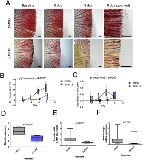
Anti-angiogenic drug SU5416 reduces caudal fin regeneration in aged fish. Caudal fin angiogenesis assay with old turquoise killifish (aged 32 weeks) and control or SU5416 treatment. (A) Microscopy images of caudal fins of turquoise killifish during caudal fin regeneration assay n=6 for DMSO and n=8 for SU5416. Scale bar: 1 mm. (B) Quantification of total regeneration of caudal fin. (C) Quantification of thickness of regenerating area. (D) Measurement of blood vessel density in histological sections. (E,F) qPCR measurements of mRNA expression of vascular biomarkers cd34 (E) and vegfa (F). n=7 for both groups. Statistical analysis in C and D is two-way ANOVA (treatment and time as factors, all time points used in analysis) and in D, E and F Mann–Whitney U-test. |
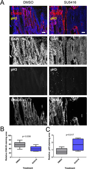
Anti-angiogenic drug SU5416 alters cellular response during turquoise killifish caudal fin regeneration. Samples were obtained from turquoise killifish (age 32 weeks) caudal fin regeneration assay and processed to histological sections. (A) Image of immunostaining for proliferation (pH3), collagen (CNA35) and nuclei (DAPI). Scale bar: 0.1 mm. (B) Quantification of collagen content stained by CNA-35 probe. (C) Quantification of cell proliferation marker phospho-histone 3 (pH3). n=7 in both groups. |