FIGURE SUMMARY
Title

Molecular Actors of Inflammation and Their Signaling Pathways: Mechanistic Insights from Zebrafish

Authors
Leiba, J., Özbilgiç, R., Hernández, L., Demou, M., Lutfalla, G., Yatime, L., Nguyen-Chi, M.
Source
Full text @ Biology (Basel)

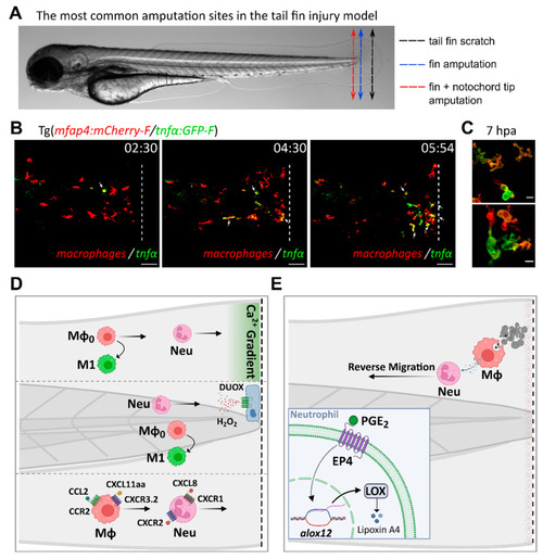
Inflammatory mediators and signaling pathways regulating leukocyte recruitment and activation during sterile injury, as inferred from the tail fin injury model. (A). The most common amputation/wounding sites used in the tail fin injury model are highlighted on a representative transmitted light image of a 3 dpf larva. Black dotted arrow: scratch on the tail fin tip. Blue dotted arrow: transection of the fin tip only, just before the notochord. Red dotted arrow: transection of the fin and the tip of the notochord. (B). Time-course of macrophage recruitment and activation following tail fin transection (intact notochord). Real-time imaging of the fin fold of a 3 dpf amputated larva from the Tg(mfap4:mCherry-F/tnfα:GFP-F) line, using confocal microscopy (unpublished data by Nguyen-Chi M. et al). In this line, macrophages and TNFα-expressing cells are labelled with red and green fluorescence, respectively. The representative frames show macrophage recruitment and activation toward an M1 phenotype at the wound, as previously observed by Sipka et al [53]. M1 macrophages, i.e., macrophages expressing TNFα, are indicated with white arrows. Time after amputation is indicated in hour:min. Dotted lines show the wound margin. Scale bar: 50 µm. (C). Representative images of activated macrophages at high magnification at 7 h post-amputation (unpublished data by Nguyen-Chi M. et al). Scale bar: 10 µm. (D). Tail wounding with fin amputation only. Upper part: fin wounding triggers instantaneous calcium flashes that are required for the recruitment of both neutrophils and macrophages. Ca2+ signaling is also important for macrophage polarization toward M1-like phenotype [53]. Middle part: reactive oxygen species H2O2 induces the recruitment of neutrophils at the wound site [112,113]. ROS are also required for macrophage M1-like activation [53]. Lower part: chemokines are essential for leukocyte recruitment to injury sites. Neutrophils migrate toward the wound via the CXCL8/CXCR1 axis while the CXCL8/CXCR2 axis promotes their dispersal [81,304]. By contrast, macrophage recruitment relies on CCL2/CCR2 and CXCL11aa/CXCR3.2 axes [221,225]. (E). Tail wounding with both fin and notochord tip amputation. Macrophage uptake of apoptotic cells triggers the resolution of inflammation by the production of PGE2 [285]. Mechanistically, PGE2 acts via EP4 receptors present on neutrophils, stimulating LOX expression. LOX activity then induces a lipid mediator switch that activates lipoxin production. Mϕ: macrophage. Neu: neutrophil.

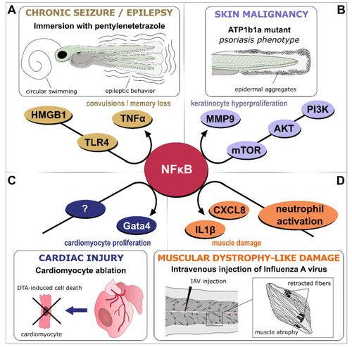
NFκB-dependent signaling as a central hub in inflammation. Various models of inflammatory-driven pathologies generated in larvae or adult zebrafish have identified NFκB signaling as part of the disease onset, progression, or recovery process. (A). In an adult model of epileptic seizure induced by pentylenetetrazole, the HMGB1/TLR4/NFκB pathway generates deleterious inflammation that results in a convulsive behavior [310]. (B). In larvae, atp1b1a mutation results in psoriasis-like phenotype and skin malignancy, through activation of the PI3K-AKT-mTORC1-NFκB-MMP9 pathway, which promotes keratinocytes overgrowth and invasiveness of epidermal layers [314]. (C). In contrast to these deleterious effects, NFκB pro-inflammatory signaling is required for cardiac regeneration as exemplified in an adult model of genetic cardiomyocyte ablation where NFκB-dependent upregulation of gata4-controled genes permits cardiomyocyte proliferation and epicardial infiltration [315]. (D). Infection of zebrafish larvae by Influenza A virus (IAV) induces a neutrophil-dependent activation of NFκB in skeletal muscles, upregulating pro-inflammatory cytokines such as IL1β and CXCL8 and generating sustained muscle damage that mimics Duchenne muscular dystrophy [316].

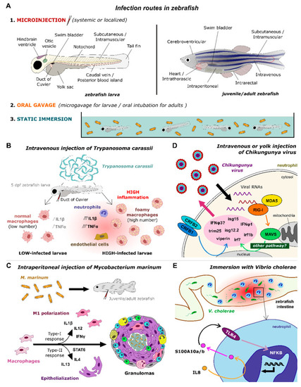
Signaling pathways unraveled by infection models in zebrafish. (A). Infection routes used in zebrafish models include micro-injection in various organs/tissues, oral gavage, and immersion of the pathogens in the fish water, both for larvae and adult zebrafish. (B). A model of infection by Trypanosoma carassii through intravenous injection in 5 dpf larvae revealed two distinct macrophage pro-inflammatory phenotypes depending on the level of infection, high-infected larvae producing foamy macrophages with strong pro-inflammatory response and incapacity to control the parasitemia [199]. (C). A model of infection by Mycobacterium marinum through intraperitoneal injection in juvenile or adult zebrafish showed that both M1- and M2-type macrophage responses are needed for mycobacterial pathogenesis and formation of granulomas [338]. (D). A model of infection by Chikungunya virus through intravenous or yolk injection in larvae uncovered the interferon-dependent antiviral immune response elicited by neutrophils [77]. (E). An adult model of infection by Vibrio cholerae through immersion evidenced the NFκB-dependent recruitment of leukocytes in the intestine through activation of an IL8/S100/TLR4 pathway [339].

Acknowledgments
This image is the copyrighted work of the attributed author or publisher, and ZFIN has permission only to display this image to its users. Additional permissions should be obtained from the applicable author or publisher of the image. Full text @ Biology (Basel)