FIGURE SUMMARY
Title

Single-cell transcriptomes and runx2b-/- mutants reveal the genetic signatures of intermuscular bone formation in zebrafish

Authors
Nie, C.H., Wan, S.M., Chen, Y.L., Huysseune, A., Wu, Y.M., Zhou, J.J., Hilsdorf, A.W.S., Wang, W.M., Witten, P.E., Lin, Q., Gao, Z.X.
Source
Full text @ Natl Sci Rev

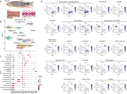
Global patterns of single-cell expression profiles of the tissue of origin of intermuscular bones (IBs) in wild-type zebrafish, and identification of cell types. (A) Pattern of tissue collection for scRNA-seq. (B) Uniform manifold approximation and projection (UMAP) visualization of 18 subsets from 13075 cells from IB-containing muscle in wild-type zebrafish. (C) Dot plots showing the expression of curated feature genes in 18 subsets. Dot sizes represent the proportion of cells expressing a specific gene in the indicated subset and color bars represent the gene expression levels. (D) UMAP visualization of the expression of curated feature genes for tenocyte and osteoblast identification. The color bar represents the gene expression levels.

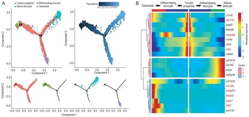
Differentiation trajectory construction of tendons and intermuscular bones, and screening of key regulators for IB formation. (A) Diffusion map visualization of the tendon and IB trajectories simulated by monocle2 across tendon progenitors, mature tenocytes, differentiating tenocytes and osteoblasts on the left. The corresponding diffusion pseudo-time is indicated in the bottom-left frame. A diffusion map visualization of each cluster is shown on the right. (B) Heatmap of the gene expression (smoothed over three adjacent cells) in subsets ordered by the pseudo-time of mature tenocytes and osteoblasts, as in (A); some curated genes are shown.

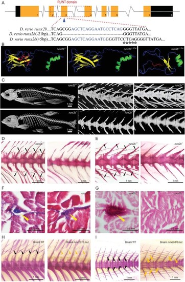
Gene structure and phenotypic characteristics of runx2b+/+ and runx2b−/−fish. (A) runx2b gene structure and RUNT domain characteristics. CRISPR-Cas9 target sequences are marked as blue letters in the DNA sequences. Deleted nucleotides are shown by ‘-’ and inserted sequences are marked as ‘*’ in runx2b−/− zebrafish. (B) The runx2b protein structure. The β-sheet domain changed in runx2b−/− zebrafish. The red area indicates the missing or changed amino acids in runx2b−/− zebrafish compared with those in runx2b+/+ zebrafish. The α-helix is indicated in green; β-sheet is indicated in yellow; random coil is indicated in blue. (C) Micro-CT scan with a resolution under 6 μm showing overall skeletal structures. Regions of vertebrae 9–14 and 19–23 were scanned with micro-CT under 4 μm resolution to show details of the IBs. (D and E) Alizarin red S staining of the abdominal and caudal region. IBs (arrowheads) are lost in runx2b−/− zebrafish. (F) Masson's trichrome staining of transverse paraffin sections. The general tissue organization is maintained in runx2b−/− zebrafish compared to that in runx2b+/+ zebrafish. (G) Transverse paraffin sections of the area normally containing IBs (arrowheads) stained with Alizarin red S, indicated the loss of mineralized IBs in runx2b−/− zebrafish. (H and I) IBs phenotype of runx2b F0 generation mutants in blunt snout bream. The IBs are partially lost in the tail and back parts of the runx2b F0 generation mutants. The black arrowhead indicates mineralized IBs, while yellow arrowhead indicates incomplete mineralized IBs.

Comparison of characteristics of other bones, swimming performance and muscle nutrient content between runx2b+/+ and runx2b−/− zebrafish. (A and B) Tissue mineral density (TMD) value of six distinctive bone elements. (C and D) Quantification of muscle and fat volumes. (E and F) Body weight and body length. (G) Muscle fiber area of tail muscles, calculated based on hematoxylin and eosin (HE)-stained sections. (H) Average swimming velocity. (I) Fatty acid contents in the muscle tissue of zebrafish at 90 dpf. (J) Amino acid contents in the muscle tissue of zebrafish at 90 dpf. ns, P > 0.05; *, P < 0.05.

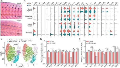
Comparative transcriptome analysis of runx2b+/+ and runx2b−/− zebrafish, and overexpression and knockdown analysis of runx2b. (A) In situ hybridization of runx2b in the tissue of origin of IBs (IBs, shown with arrowheads). The expression of runx2b was significantly reduced in runx2b−/− zebrafish. (B) UMAP visualization of tendon progenitors, mature tenocytes, differentiating tenocytes and osteoblasts in runx2b+/+ and runx2b−/−zebrafish. The ratios of osteoblasts to the total number of cells in runx2b+/+ and runx2b−/− zebrafish were 2.44% (340 cells) and 0.31% (34 cells), respectively. (C) Violin plots showing expression of differentially expressed genes in each cluster related to IB formation between runx2b+/+ and runx2b−/−zebrafish. (D, E) Overexpression and knockdown analysis of runx2b in IB cells of M. amblycephala. The expression levels of genes related to osteoblasts were detected by qRT-PCR. ANOVA (analysis of variance) was used to test the differences in expression. ns, P > 0.05; *, P < 0.05; **, P < 0.01; ***, P < 0.001.

Acknowledgments
This image is the copyrighted work of the attributed author or publisher, and ZFIN has permission only to display this image to its users. Additional permissions should be obtained from the applicable author or publisher of the image. Full text @ Natl Sci Rev